4LIX | pdb_00004lix

Crystal structure of ent-copalyl diphosphate synthase from Arabidopsis thaliana in complex with (S)-15-aza-14,15-dihydrogeranylgeranyl thiolodiphosphate at 1.55 A resolution


Experimental Data Snapshot

  • Method: X-RAY DIFFRACTION
  • Resolution: 1.55 Å
  • R-Value Free: 
    0.196 (Depositor), 0.192 (DCC) 
  • R-Value Work: 
    0.169 (Depositor), 0.165 (DCC) 
  • R-Value Observed: 
    0.170 (Depositor) 

Starting Model: experimental
View more details

wwPDB Validation 3D Report Full Report

Validation slider image for 4LIX

Ligand Structure Quality Assessment 


This is version 1.4 of the entry. See complete history

Literature

1.55 angstrom -resolution structure of ent-copalyl diphosphate synthase and exploration of general acid function by site-directed mutagenesis.

Koksal, M.Potter, K.Peters, R.J.Christianson, D.W.

(2013) Biochim Biophys Acta 1840: 184-190

  • DOI: https://doi.org/10.1016/j.bbagen.2013.09.004
  • Primary Citation Related Structures: 
    4LIX

  • PubMed Abstract: 

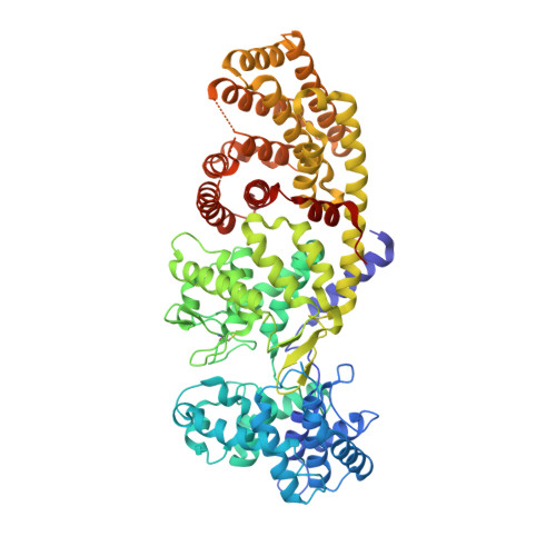
    The diterpene cyclase ent-copalyl diphosphate synthase (CPS) catalyzes the first committed step in the biosynthesis of gibberellins. The previously reported 2.25Å resolution crystal structure of CPS complexed with (S)-15-aza-14,15-dihydrogeranylgeranyl thiolodiphosphate (1) established the αβγ domain architecture, but ambiguities regarding substrate analog binding remained. Use of crystallization additives yielded CPS crystals diffracting to 1.55Å resolution. Additionally, active site residues that hydrogen bond with D379, either directly or through hydrogen bonded water molecules, were probed by mutagenesis. This work clarifies structure-function relationships that were ambiguous in the lower resolution structure. Well-defined positions for the diphosphate group and tertiary ammonium cation of 1, as well as extensive solvent structure, are observed. Two channels involving hydrogen bonded solvent and protein residues lead to the active site, forming hydrogen bonded "proton wires" that link general acid D379 with bulk solvent. These proton wires may facilitate proton transfer with the general acid during catalysis. Activity measurements made with mutant enzymes indicate that N425, which donates a hydrogen bond directly to D379, and T421, which hydrogen bonds with D379 through an intervening solvent molecule, help orient D379 for catalysis. Residues involved in hydrogen bonds with the proton wire, R340 and D503, are also important. Finally, conserved residue E211, which is located near the diphosphate group of 1, is proposed to be a ligand to Mg(2+) required for optimal catalytic activity. This work establishes structure-function relationships for class II terpenoid cyclases.


  • Organizational Affiliation
    • Roy and Diana Vagelos Laboratories, Department of Chemistry, University of Pennsylvania, Philadelphia, PA 19104-6323, USA.

Macromolecule Content 

  • Total Structure Weight: 85.51 kDa 
  • Atom Count: 6,360 
  • Modeled Residue Count: 678 
  • Deposited Residue Count: 727 
  • Unique protein chains: 1

Macromolecules

Find similar proteins by:|  3D Structure
Entity ID: 1
MoleculeChains  Sequence LengthOrganismDetailsImage
Ent-copalyl diphosphate synthase, chloroplastic727Arabidopsis thalianaMutation(s): 0 
Gene Names: ABC33At4g02780CPSCPS1GA1T5J8.9TPSGA1
EC: 5.5.1.13
UniProt
Find proteins for Q38802 (Arabidopsis thaliana)
Explore Q38802 
Go to UniProtKB:  Q38802
Entity Groups
Sequence Clusters30% Identity50% Identity70% Identity90% Identity95% Identity100% Identity
UniProt GroupQ38802
Sequence Annotations
Expand
Reference Sequence

Small Molecules

Ligands 3 Unique
IDChains Name / Formula / InChI Key2D Diagram3D Interactions
AG8

Query on AG8


Download:Ideal Coordinates CCD File
B [auth A]S-[(2E,6E,10E)-14-(dimethylamino)-3,7,11-trimethyltetradeca-2,6,10-trien-1-yl] trihydrogen thiodiphosphate
C19 H37 N O6 P2 S
WNRLOKILDUQQLN-WNWLUAIZSA-N
PO4

Query on PO4


Download:Ideal Coordinates CCD File
C [auth A]PHOSPHATE ION
O4 P
NBIIXXVUZAFLBC-UHFFFAOYSA-K
GOL

Query on GOL


Download:Ideal Coordinates CCD File
D [auth A]GLYCEROL
C3 H8 O3
PEDCQBHIVMGVHV-UHFFFAOYSA-N
Modified Residues  1 Unique
IDChains TypeFormula2D DiagramParent
CSO
Query on CSO
A
L-PEPTIDE LINKINGC3 H7 N O3 SCYS

Experimental Data & Validation

Experimental Data

  • Method: X-RAY DIFFRACTION
  • Resolution: 1.55 Å
  • R-Value Free:  0.196 (Depositor), 0.192 (DCC) 
  • R-Value Work:  0.169 (Depositor), 0.165 (DCC) 
  • R-Value Observed: 0.170 (Depositor) 
Space Group: P 21 21 21
Unit Cell:
Length ( Å )Angle ( ˚ )
a = 130.177α = 90
b = 51.393β = 90
c = 114.308γ = 90
Software Package:
Software NamePurpose
CBASSdata collection
PHENIXmodel building
PHENIXrefinement
HKL-2000data reduction
HKL-2000data scaling
PHENIXphasing

Structure Validation

View Full Validation Report


Ligand Structure Quality Assessment 


Entry History 

Deposition Data

Revision History  (Full details and data files)

  • Version 1.0: 2013-10-02
    Type: Initial release
  • Version 1.1: 2013-10-30
    Changes: Database references
  • Version 1.2: 2023-09-20
    Changes: Data collection, Database references, Derived calculations, Refinement description
  • Version 1.3: 2023-12-06
    Changes: Data collection
  • Version 1.4: 2024-11-06
    Changes: Structure summary