4LH7 | pdb_00004lh7

Crystal structure of a LigA inhibitor


Experimental Data Snapshot

  • Method: X-RAY DIFFRACTION
  • Resolution: 1.90 Å
  • R-Value Free: 
    0.259 (Depositor), 0.267 (DCC) 
  • R-Value Work: 
    0.225 (Depositor), 0.233 (DCC) 
  • R-Value Observed: 
    0.226 (Depositor) 

wwPDB Validation 3D Report Full Report

Validation slider image for 4LH7

Ligand Structure Quality Assessment 


This is version 1.3 of the entry. See complete history

Literature

Identification through structure-based methods of a bacterial NAD(+)-dependent DNA ligase inhibitor that avoids known resistance mutations.

Murphy-Benenato, K.Wang, H.McGuire, H.M.Davis, H.E.Gao, N.Prince, D.B.Jahic, H.Stokes, S.S.Boriack-Sjodin, P.A.

(2014) Bioorg Med Chem Lett 24: 360-366

  • DOI: https://doi.org/10.1016/j.bmcl.2013.11.007
  • Primary Citation Related Structures: 
    4LH6, 4LH7

  • PubMed Abstract: 

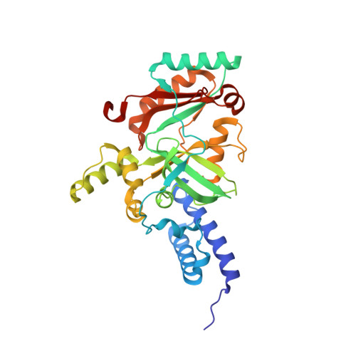
    In an attempt to identify novel inhibitors of NAD(+)-dependent DNA ligase (LigA) that are not affected by a known resistance mutation in the adenosine binding pocket, a detailed analysis of the binding sites of a variety of bacterial ligases was performed. This analysis revealed several similarities to the adenine binding region of kinases, which enabled a virtual screen of known kinase inhibitors. From this screen, a thienopyridine scaffold was identified that was shown to inhibit bacterial ligase. Further characterization through structure and enzymology revealed the compound was not affected by a previously disclosed resistance mutation in Streptococcus pneumoniae LigA, Leu75Phe. A subsequent medicinal chemistry program identified substitutions that resulted in an inhibitor with moderate activity across various Gram-positive bacterial LigA enzymes.


  • Organizational Affiliation
    • Department of Chemistry, Infection Innovative Medicines, AstraZeneca R&D Boston, 35 Gatehouse Dr., Waltham, MA 02451, United States. Electronic address: kerry.benenato@astrazeneca.com.

Macromolecule Content 

  • Total Structure Weight: 37.48 kDa 
  • Atom Count: 2,820 
  • Modeled Residue Count: 321 
  • Deposited Residue Count: 323 
  • Unique protein chains: 1

Macromolecules

Find similar proteins by:|  3D Structure
Entity ID: 1
MoleculeChains  Sequence LengthOrganismDetailsImage
DNA ligase323Enterococcus faecalis V583Mutation(s): 0 
Gene Names: EF_0722ligA
EC: 6.5.1.2
UniProt
Find proteins for Q837V6 (Enterococcus faecalis (strain ATCC 700802 / V583))
Explore Q837V6 
Go to UniProtKB:  Q837V6
Entity Groups
Sequence Clusters30% Identity50% Identity70% Identity90% Identity95% Identity100% Identity
UniProt GroupQ837V6
Sequence Annotations
Expand
Reference Sequence

Experimental Data & Validation

Experimental Data

  • Method: X-RAY DIFFRACTION
  • Resolution: 1.90 Å
  • R-Value Free:  0.259 (Depositor), 0.267 (DCC) 
  • R-Value Work:  0.225 (Depositor), 0.233 (DCC) 
  • R-Value Observed: 0.226 (Depositor) 
Space Group: C 2 2 21
Unit Cell:
Length ( Å )Angle ( ˚ )
a = 48.838α = 90
b = 105.025β = 90
c = 136.085γ = 90
Software Package:
Software NamePurpose
CrystalCleardata collection
AMoREphasing
REFMACrefinement
d*TREKdata reduction
d*TREKdata scaling

Structure Validation

View Full Validation Report


Ligand Structure Quality Assessment 


Entry History 

Deposition Data

Revision History  (Full details and data files)

  • Version 1.0: 2013-12-25
    Type: Initial release
  • Version 1.1: 2014-01-08
    Changes: Database references
  • Version 1.2: 2021-01-13
    Changes: Derived calculations, Source and taxonomy
  • Version 1.3: 2024-02-28
    Changes: Data collection, Database references