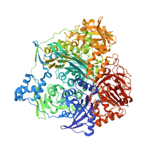
4LGY

Importance of Hydrophobic Cavities in Allosteric Regulation of Formylglycinamide Synthetase: Insight from Xenon Trapping and Statistical Coupling Analysis


Experimental Data Snapshot

  • Method: X-RAY DIFFRACTION
  • Resolution: 1.48 Å
  • R-Value Free: 0.166 
  • R-Value Work: 0.142 
  • R-Value Observed: 0.142 

Starting Model: experimental
View more details

wwPDB Validation   3D Report Full Report


Ligand Structure Quality Assessment 


This is version 1.1 of the entry. See complete history


Literature

Importance of hydrophobic cavities in allosteric regulation of formylglycinamide synthetase: insight from xenon trapping and statistical coupling analysis

Tanwar, A.S.Goyal, V.D.Choudhary, D.Panjikar, S.Anand, R.

(2013) PLoS One 8: e77781-e77781

  • DOI: https://doi.org/10.1371/journal.pone.0077781
  • Primary Citation of Related Structures:  
    4L78, 4LGY, 4MGH

  • PubMed Abstract: 

    Formylglycinamide ribonucleotide amidotransferase (FGAR-AT) is a 140 kDa bi-functional enzyme involved in a coupled reaction, where the glutaminase active site produces ammonia that is subsequently utilized to convert FGAR to its corresponding amidine in an ATP assisted fashion. The structure of FGAR-AT has been previously determined in an inactive state and the mechanism of activation remains largely unknown. In the current study, hydrophobic cavities were used as markers to identify regions involved in domain movements that facilitate catalytic coupling and subsequent activation of the enzyme. Three internal hydrophobic cavities were located by xenon trapping experiments on FGAR-AT crystals and further, these cavities were perturbed via site-directed mutagenesis. Biophysical characterization of the mutants demonstrated that two of these three voids are crucial for stability and function of the protein, although being ∼20 Å from the active centers. Interestingly, correlation analysis corroborated the experimental findings, and revealed that amino acids lining the functionally important cavities form correlated sets (co-evolving residues) that connect these regions to the amidotransferase active center. It was further proposed that the first cavity is transient and allows for breathing motion to occur and thereby serves as an allosteric hotspot. In contrast, the third cavity which lacks correlated residues was found to be highly plastic and accommodated steric congestion by local adjustment of the structure without affecting either stability or activity.


  • Organizational Affiliation

    Department of Chemistry, Indian Institute of Technology Bombay, Mumbai, India.


Macromolecules
Find similar proteins by:  (by identity cutoff)  |  3D Structure
Entity ID: 1
MoleculeChains Sequence LengthOrganismDetailsImage
Phosphoribosylformylglycinamidine synthase1,305Salmonella enterica subsp. enterica serovar Typhimurium str. LT2Mutation(s): 1 
EC: 6.3.5.3
UniProt
Find proteins for P74881 (Salmonella typhimurium (strain LT2 / SGSC1412 / ATCC 700720))
Explore P74881 
Go to UniProtKB:  P74881
Entity Groups  
Sequence Clusters30% Identity50% Identity70% Identity90% Identity95% Identity100% Identity
UniProt GroupP74881
Sequence Annotations
Expand
  • Reference Sequence
Small Molecules
Ligands 6 Unique
IDChains Name / Formula / InChI Key2D Diagram3D Interactions
ADP
Query on ADP

Download Ideal Coordinates CCD File 
B [auth A]ADENOSINE-5'-DIPHOSPHATE
C10 H15 N5 O10 P2
XTWYTFMLZFPYCI-KQYNXXCUSA-N
SO4
Query on SO4

Download Ideal Coordinates CCD File 
F [auth A]
G [auth A]
H [auth A]
I [auth A]
J [auth A]
F [auth A],
G [auth A],
H [auth A],
I [auth A],
J [auth A],
K [auth A],
L [auth A],
M [auth A],
N [auth A],
O [auth A],
P [auth A],
Q [auth A],
R [auth A],
S [auth A],
T [auth A],
U [auth A],
V [auth A],
W [auth A],
X [auth A]
SULFATE ION
O4 S
QAOWNCQODCNURD-UHFFFAOYSA-L
GOL
Query on GOL

Download Ideal Coordinates CCD File 
BA [auth A],
CA [auth A],
DA [auth A],
EA [auth A]
GLYCEROL
C3 H8 O3
PEDCQBHIVMGVHV-UHFFFAOYSA-N
ACT
Query on ACT

Download Ideal Coordinates CCD File 
AA [auth A],
Y [auth A],
Z [auth A]
ACETATE ION
C2 H3 O2
QTBSBXVTEAMEQO-UHFFFAOYSA-M
CL
Query on CL

Download Ideal Coordinates CCD File 
FA [auth A]
GA [auth A]
HA [auth A]
IA [auth A]
JA [auth A]
FA [auth A],
GA [auth A],
HA [auth A],
IA [auth A],
JA [auth A],
KA [auth A]
CHLORIDE ION
Cl
VEXZGXHMUGYJMC-UHFFFAOYSA-M
MG
Query on MG

Download Ideal Coordinates CCD File 
C [auth A]
D [auth A]
E [auth A]
LA [auth A]
MA [auth A]
C [auth A],
D [auth A],
E [auth A],
LA [auth A],
MA [auth A],
NA [auth A],
OA [auth A]
MAGNESIUM ION
Mg
JLVVSXFLKOJNIY-UHFFFAOYSA-N
Modified Residues  1 Unique
IDChains TypeFormula2D DiagramParent
CYG
Query on CYG
A
L-PEPTIDE LINKINGC8 H14 N2 O5 SCYS
Experimental Data & Validation

Experimental Data

  • Method: X-RAY DIFFRACTION
  • Resolution: 1.48 Å
  • R-Value Free: 0.166 
  • R-Value Work: 0.142 
  • R-Value Observed: 0.142 
  • Space Group: P 65
Unit Cell:
Length ( Å )Angle ( ˚ )
a = 146.329α = 90
b = 146.329β = 90
c = 140.841γ = 120
Software Package:
Software NamePurpose
REFMACrefinement
PDB_EXTRACTdata extraction
iMOSFLMdata reduction
SCALAdata scaling

Structure Validation

View Full Validation Report


Ligand Structure Quality Assessment 


Entry History 

Deposition Data

Revision History  (Full details and data files)

  • Version 1.0: 2013-12-11
    Type: Initial release
  • Version 1.1: 2023-11-08
    Changes: Data collection, Database references, Derived calculations, Refinement description