4LG2

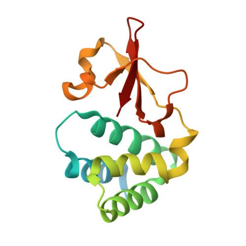
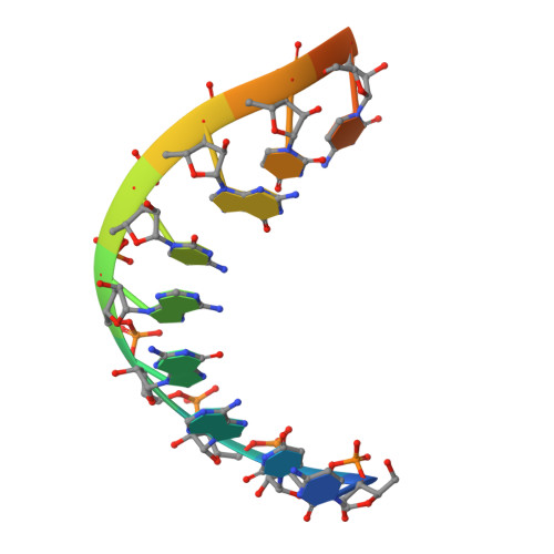
Crystal structure of Reston Ebola virus VP35 RNA binding domain bound to 12-bp dsRNA


Experimental Data Snapshot

  • Method: X-RAY DIFFRACTION
  • Resolution: 2.70 Å
  • R-Value Free: 0.262 
  • R-Value Work: 0.209 
  • R-Value Observed: 0.211 

Starting Model: experimental
View more details

wwPDB Validation   3D Report Full Report


This is version 1.5 of the entry. See complete history


Literature

Ebolavirus VP35 coats the backbone of double-stranded RNA for interferon antagonism.

Bale, S.Julien, J.P.Bornholdt, Z.A.Krois, A.S.Wilson, I.A.Saphire, E.O.

(2013) J Virol 87: 10385-10388

  • DOI: https://doi.org/10.1128/JVI.01452-13
  • Primary Citation of Related Structures:  
    4LG2

  • PubMed Abstract: 

    Recognition of viral double-stranded RNA (dsRNA) activates interferon production and immune signaling in host cells. Crystal structures of ebolavirus VP35 show that it caps dsRNA ends to prevent sensing by pattern recognition receptors such as RIG-I. In contrast, structures of marburgvirus VP35 show that it primarily coats the dsRNA backbone. Here, we demonstrate that ebolavirus VP35 also coats the dsRNA backbone in solution, although binding to the dsRNA ends probably constitutes the initial binding event.


  • Organizational Affiliation

    Department of Immunology and Microbial Science, The Scripps Research Institute, La Jolla, California, USA.


Macromolecules

Find similar proteins by:  (by identity cutoff)  |  3D Structure
Entity ID: 1
MoleculeChains Sequence LengthOrganismDetailsImage
Polymerase cofactor
A, B, C, D
146Reston ebolavirus - Reston (1989)Mutation(s): 0 
Gene Names: VP35REBOVgp2
UniProt
Find proteins for Q8JPY0 (Reston ebolavirus (strain Reston-89))
Explore Q8JPY0 
Go to UniProtKB:  Q8JPY0
Entity Groups  
Sequence Clusters30% Identity50% Identity70% Identity90% Identity95% Identity100% Identity
UniProt GroupQ8JPY0
Sequence Annotations
Expand
  • Reference Sequence

Find similar nucleic acids by:  Sequence   |   3D Structure  

Entity ID: 2
MoleculeChains LengthOrganismImage
dsRNAE,
F,
G [auth J],
H [auth I]
12N/A
Sequence Annotations
Expand
  • Reference Sequence
Experimental Data & Validation

Experimental Data

  • Method: X-RAY DIFFRACTION
  • Resolution: 2.70 Å
  • R-Value Free: 0.262 
  • R-Value Work: 0.209 
  • R-Value Observed: 0.211 
  • Space Group: P 21 21 21
Unit Cell:
Length ( Å )Angle ( ˚ )
a = 55.09α = 90
b = 72.871β = 90
c = 175.551γ = 90
Software Package:
Software NamePurpose
ADSCdata collection
PHENIXmodel building
PHENIXrefinement
d*TREKdata reduction
d*TREKdata scaling
PHENIXphasing

Structure Validation

View Full Validation Report


Entry History 

Deposition Data

Revision History  (Full details and data files)

  • Version 1.0: 2013-07-10
    Type: Initial release
  • Version 1.1: 2013-07-17
    Changes: Database references
  • Version 1.2: 2013-11-27
    Changes: Database references
  • Version 1.3: 2017-06-14
    Changes: Structure summary
  • Version 1.4: 2018-01-24
    Changes: Structure summary
  • Version 1.5: 2023-09-20
    Changes: Data collection, Database references, Refinement description