4LFE | pdb_00004lfe

Crystal structure of geranylgeranyl diphosphate synthase sub1274 (target efi-509455) from streptococcus uberis 0140j with bound magnesium and isopentyl diphosphate, partially liganded complex;


Experimental Data Snapshot

  • Method: X-RAY DIFFRACTION
  • Resolution: 1.95 Å
  • R-Value Free: 
    0.236 (Depositor), 0.241 (DCC) 
  • R-Value Work: 
    0.207 (Depositor), 0.214 (DCC) 
  • R-Value Observed: 
    0.208 (Depositor) 

Starting Model: experimental
View more details

wwPDB Validation 3D Report Full Report

Validation slider image for 4LFE

Ligand Structure Quality Assessment 


This is version 1.1 of the entry. See complete history

Literature

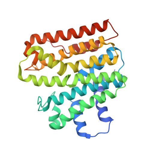
Crystal Structure of Geranylgeranyl Diphosphate Synthase from Streptococcus Uberis 0140J

Patskovsky, Y.Toro, R.Bhosle, R.Hillerich, B.Seidel, R.D.Washington, E.Scott Glenn, A.Chowdhury, S.Evans, B.Hammonds, J.Imker, H.J.Al Obaidi, N.Stead, M.Love, J.Poulter, C.D.Gerlt, J.A.Almo, S.C.

To be published.

Macromolecule Content 

  • Total Structure Weight: 65.46 kDa 
  • Atom Count: 4,662 
  • Modeled Residue Count: 562 
  • Deposited Residue Count: 580 
  • Unique protein chains: 1

Macromolecules

Find similar proteins by:|  3D Structure
Entity ID: 1
MoleculeChains  Sequence LengthOrganismDetailsImage
Geranylgeranyl diphosphate synthase
A, B
290Streptococcus uberis 0140JMutation(s): 0 
Gene Names: fpsSUB1274
EC: 2.5.1.10
UniProt
Find proteins for B9DUT7 (Streptococcus uberis (strain ATCC BAA-854 / 0140J))
Explore B9DUT7 
Go to UniProtKB:  B9DUT7
Entity Groups
Sequence Clusters30% Identity50% Identity70% Identity90% Identity95% Identity100% Identity
UniProt GroupB9DUT7
Sequence Annotations
Expand
Reference Sequence

Small Molecules

Ligands 2 Unique
IDChains Name / Formula / InChI Key2D Diagram3D Interactions
IPE

Query on IPE


Download:Ideal Coordinates CCD File
C [auth A],
F [auth B],
G [auth B]
3-METHYLBUT-3-ENYL TRIHYDROGEN DIPHOSPHATE
C5 H12 O7 P2
NUHSROFQTUXZQQ-UHFFFAOYSA-N
MG

Query on MG


Download:Ideal Coordinates CCD File
D [auth A],
E [auth A],
H [auth B],
I [auth B],
J [auth B]
MAGNESIUM ION
Mg
JLVVSXFLKOJNIY-UHFFFAOYSA-N

Experimental Data & Validation

Experimental Data

  • Method: X-RAY DIFFRACTION
  • Resolution: 1.95 Å
  • R-Value Free:  0.236 (Depositor), 0.241 (DCC) 
  • R-Value Work:  0.207 (Depositor), 0.214 (DCC) 
  • R-Value Observed: 0.208 (Depositor) 
Space Group: P 21 21 21
Unit Cell:
Length ( Å )Angle ( ˚ )
a = 70.798α = 90
b = 75.789β = 90
c = 124.62γ = 90
Software Package:
Software NamePurpose
PHASERphasing
REFMACrefinement
HKL-2000data reduction
HKL-2000data scaling

Structure Validation

View Full Validation Report


Ligand Structure Quality Assessment 


Entry History 

Revision History  (Full details and data files)

  • Version 1.0: 2013-07-10
    Type: Initial release
  • Version 1.1: 2023-09-20
    Changes: Data collection, Database references, Derived calculations, Refinement description