4LE6 | pdb_00004le6

Crystal structure of the phosphotriesterase OPHC2 from Pseudomonas pseudoalcaligenes


Experimental Data Snapshot

  • Method: X-RAY DIFFRACTION
  • Resolution: 2.10 Å
  • R-Value Free: 
    0.210 (Depositor), 0.214 (DCC) 
  • R-Value Work: 
    0.173 (Depositor), 0.178 (DCC) 
  • R-Value Observed: 
    0.175 (Depositor) 

Starting Model: experimental
View more details

wwPDB Validation 3D Report Full Report

Validation slider image for 4LE6

This is version 1.3 of the entry. See complete history

Literature

Structural and Enzymatic Characterization of the Phosphotriesterase OPHC2 from Pseudomonas pseudoalcaligenes.

Gotthard, G.Hiblot, J.Gonzalez, D.Elias, M.Chabriere, E.

(2013) PLoS One 8: e77995-e77995

  • DOI: https://doi.org/10.1371/journal.pone.0077995
  • Primary Citation Related Structures: 
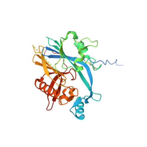
    4LE6

  • PubMed Abstract: 

    Organophosphates (OPs) are neurotoxic compounds for which current methods of elimination are unsatisfactory; thus bio-remediation is considered as a promising alternative. Here we provide the structural and enzymatic characterization of the recently identified enzyme isolated from Pseudomonas pseudoalcaligenes dubbed OPHC2. OPHC2 belongs to the metallo-β-lactamase superfamily and exhibits an unusual thermal resistance and some OP degrading abilities. The X-ray structure of OPHC2 has been solved at 2.1 Å resolution. The enzyme is roughly globular exhibiting a αβ/βα topology typical of the metallo-β-lactamase superfamily. Several structural determinants, such as an extended dimerization surface and an intramolecular disulfide bridge, common features in thermostable enzymes, are consistent with its high Tm (97.8°C). Additionally, we provide the enzymatic characterization of OPHC2 against a wide range of OPs, esters and lactones. OPHC2 possesses a broad substrate activity spectrum, since it hydrolyzes various phosphotriesters, esters, and a lactone. Because of its organophosphorus hydrolase activity, and given its intrinsic thermostability, OPHC2 is an interesting candidate for the development of an OPs bio-decontaminant. Its X-ray structure shed light on its active site, and provides key information for the understanding of the substrate binding mode and catalysis.


  • Organizational Affiliation
    • URMITE UMR CNRS-IRD 6236, IFR48, Faculté de Médecine et de Pharmacie, Université de la Méditerranée, Marseille, France.

Macromolecule Content 

  • Total Structure Weight: 176.59 kDa 
  • Atom Count: 10,449 
  • Modeled Residue Count: 1,268 
  • Deposited Residue Count: 1,620 
  • Unique protein chains: 1

Macromolecules

Find similar proteins by:|  3D Structure
Entity ID: 1
MoleculeChains  Sequence LengthOrganismDetailsImage
Organophosphorus hydrolase
A, B, C, D, E
324Ectopseudomonas oleovoransMutation(s): 0 
Gene Names: ophc2
EC: 3.1.8.1
UniProt
Find proteins for Q5W503 (Ectopseudomonas oleovorans)
Explore Q5W503 
Go to UniProtKB:  Q5W503
Entity Groups
Sequence Clusters30% Identity50% Identity70% Identity90% Identity95% Identity100% Identity
UniProt GroupQ5W503
Sequence Annotations
Expand
Reference Sequence

Small Molecules

Ligands 4 Unique
IDChains Name / Formula / InChI Key2D Diagram3D Interactions
PGE

Query on PGE


Download:Ideal Coordinates CCD File
I [auth A]TRIETHYLENE GLYCOL
C6 H14 O4
ZIBGPFATKBEMQZ-UHFFFAOYSA-N
GOL

Query on GOL


Download:Ideal Coordinates CCD File
F [auth A]
G [auth A]
M [auth B]
N [auth B]
O [auth B]
F [auth A],
G [auth A],
M [auth B],
N [auth B],
O [auth B],
S [auth C],
T [auth C],
X [auth D]
GLYCEROL
C3 H8 O3
PEDCQBHIVMGVHV-UHFFFAOYSA-N
ZN

Query on ZN


Download:Ideal Coordinates CCD File
AA [auth E]
BA [auth E]
J [auth A]
K [auth A]
Q [auth B]
AA [auth E],
BA [auth E],
J [auth A],
K [auth A],
Q [auth B],
R [auth B],
V [auth C],
W [auth C],
Y [auth D],
Z [auth D]
ZINC ION
Zn
PTFCDOFLOPIGGS-UHFFFAOYSA-N
EDO

Query on EDO


Download:Ideal Coordinates CCD File
H [auth A],
L [auth A],
P [auth B],
U [auth C]
1,2-ETHANEDIOL
C2 H6 O2
LYCAIKOWRPUZTN-UHFFFAOYSA-N

Experimental Data & Validation

Experimental Data

  • Method: X-RAY DIFFRACTION
  • Resolution: 2.10 Å
  • R-Value Free:  0.210 (Depositor), 0.214 (DCC) 
  • R-Value Work:  0.173 (Depositor), 0.178 (DCC) 
  • R-Value Observed: 0.175 (Depositor) 
Space Group: C 1 2 1
Unit Cell:
Length ( Å )Angle ( ˚ )
a = 109.91α = 90
b = 63.85β = 101.85
c = 221.26γ = 90
Software Package:
Software NamePurpose
PHASERphasing
PHENIXrefinement
XDSdata reduction
XSCALEdata scaling

Structure Validation

View Full Validation Report


Entry History 

Deposition Data

Revision History  (Full details and data files)

  • Version 1.0: 2013-11-13
    Type: Initial release
  • Version 1.1: 2013-12-04
    Changes: Database references
  • Version 1.2: 2023-09-20
    Changes: Data collection, Database references, Derived calculations, Refinement description
  • Version 1.3: 2024-11-27
    Changes: Structure summary