4LBF | pdb_00004lbf

Crystal structure of HUMAN ALPHA-DEFENSIN 1 (HNP1) I20A/L25A mutant


Experimental Data Snapshot

  • Method: X-RAY DIFFRACTION
  • Resolution: 1.70 Å
  • R-Value Free: 
    0.231 (Depositor), 0.235 (DCC) 
  • R-Value Work: 
    0.185 (Depositor), 0.188 (DCC) 
  • R-Value Observed: 
    0.188 (Depositor) 

Starting Model: experimental
View more details

wwPDB Validation 3D Report Full Report

Validation slider image for 4LBF

This is version 1.2 of the entry. See complete history

Literature

Single, Double and Quadruple Alanine Substitutions at Oligomeric Interfaces Identify Hydrophobicity as the Key Determinant of Human Neutrophil Alpha Defensin HNP1 Function.

Zhao, L.Tolbert, W.D.Ericksen, B.Zhan, C.Wu, X.Yuan, W.Li, X.Pazgier, M.Lu, W.

(2013) PLoS One 8: e78937-e78937

  • DOI: https://doi.org/10.1371/journal.pone.0078937
  • Primary Citation Related Structures: 
    4LB1, 4LB7, 4LBB, 4LBF

  • PubMed Abstract: 

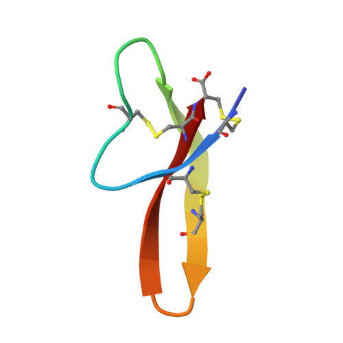
    HNP1 is a human alpha defensin that forms dimers and multimers governed by hydrophobic residues, including Tyr¹⁶, Ile²⁰, Leu²⁵, and Phe²⁸. Previously, alanine scanning mutagenesis identified each of these residues and other hydrophobic residues as important for function. Here we report further structural and functional studies of residues shown to interact with one another across oligomeric interfaces: I20A-HNP1 and L25A-HNP1, plus the double alanine mutants I20A/L25A-HNP1 and Y16A/F28A-HNP1, and the quadruple alanine mutant Y16A/I20A/L25A/F28A-HNP1. We tested binding to HIV-1 gp120 and HNP1 by surface plasmon resonance, binding to HIV-1 gp41 and HNP1 by fluorescence polarization, inhibition of anthrax lethal factor, and antibacterial activity using the virtual colony count assay. Similar to the previously described single mutant W26A-HNP1, the quadruple mutant displayed the least activity in all functional assays, followed by the double mutant Y16A/F28A-HNP1. The effects of the L25A and I20A single mutations were milder than the double mutant I20A/L25A-HNP1. Crystallographic studies confirmed the correct folding and disulfide pairing, and depicted an array of dimeric and tetrameric structures. These results indicate that side chain hydrophobicity is the critical factor that determines activity at these positions.


  • Organizational Affiliation
    • Translational Medicine Center, the First Affiliated Hospital, School of Medicine, Xi'an Jiaotong University, Xi'an, China ; Institute of Human Virology and Department of Biochemistry and Molecular Biology, University of Maryland School of Medicine, Baltimore, Maryland, United States of America.

Macromolecule Content 

  • Total Structure Weight: 27.04 kDa 
  • Atom Count: 2,036 
  • Modeled Residue Count: 235 
  • Deposited Residue Count: 240 
  • Unique protein chains: 1

Macromolecules

Find similar proteins by:|  3D Structure
Entity ID: 1
MoleculeChains  Sequence LengthOrganismDetailsImage
Neutrophil defensin 1
A, B, C, D, E
A, B, C, D, E, F, G, H
30Homo sapiensMutation(s): 2 
UniProt & NIH Common Fund Data Resources
Find proteins for P59665 (Homo sapiens)
Explore P59665 
Go to UniProtKB:  P59665
Entity Groups
Sequence Clusters30% Identity50% Identity70% Identity90% Identity95% Identity100% Identity
UniProt GroupP59665
Sequence Annotations
Expand
Reference Sequence

Small Molecules

Ligands 1 Unique
IDChains Name / Formula / InChI Key2D Diagram3D Interactions
GOL

Query on GOL


Download:Ideal Coordinates CCD File
I [auth D]GLYCEROL
C3 H8 O3
PEDCQBHIVMGVHV-UHFFFAOYSA-N

Experimental Data & Validation

Experimental Data

  • Method: X-RAY DIFFRACTION
  • Resolution: 1.70 Å
  • R-Value Free:  0.231 (Depositor), 0.235 (DCC) 
  • R-Value Work:  0.185 (Depositor), 0.188 (DCC) 
  • R-Value Observed: 0.188 (Depositor) 
Space Group: C 1 2 1
Unit Cell:
Length ( Å )Angle ( ˚ )
a = 75.002α = 90
b = 62.575β = 99.69
c = 42.498γ = 90
Software Package:
Software NamePurpose
HKL-2000data collection
PHASERphasing
REFMACrefinement
HKL-2000data reduction
HKL-2000data scaling

Structure Validation

View Full Validation Report


Entry History 

Deposition Data

Revision History  (Full details and data files)

  • Version 1.0: 2013-11-27
    Type: Initial release
  • Version 1.1: 2023-09-20
    Changes: Data collection, Database references, Derived calculations, Refinement description
  • Version 1.2: 2024-11-06
    Changes: Structure summary