4L4T | pdb_00004l4t

Structure of human MAIT TCR in complex with human MR1-6-FP


Experimental Data Snapshot

  • Method: X-RAY DIFFRACTION
  • Resolution: 2.00 Å
  • R-Value Free: 
    0.225 (Depositor), 0.239 (DCC) 
  • R-Value Work: 
    0.184 (Depositor), 0.195 (DCC) 
  • R-Value Observed: 
    0.186 (Depositor) 

Starting Model: experimental
View more details

wwPDB Validation 3D Report Full Report

Validation slider image for 4L4T

Ligand Structure Quality Assessment 


This is version 1.3 of the entry. See complete history

Literature

Recognition of vitamin B metabolites by mucosal-associated invariant T cells.

Patel, O.Kjer-Nielsen, L.Le Nours, J.Eckle, S.B.Birkinshaw, R.Beddoe, T.Corbett, A.J.Liu, L.Miles, J.J.Meehan, B.Reantragoon, R.Sandoval-Romero, M.L.Sullivan, L.C.Brooks, A.G.Chen, Z.Fairlie, D.P.McCluskey, J.Rossjohn, J.

(2013) Nat Commun 4: 2142-2142

  • DOI: https://doi.org/10.1038/ncomms3142
  • Primary Citation Related Structures: 
    4L4T, 4L4V

  • PubMed Abstract: 

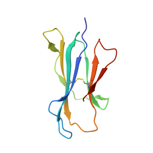
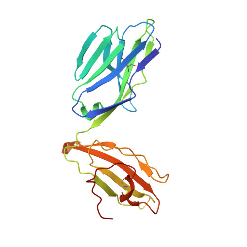
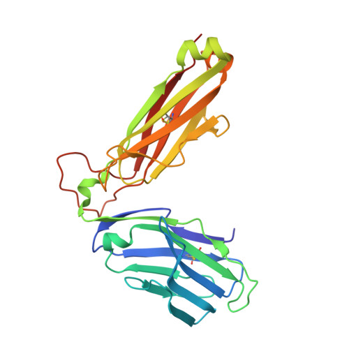
    The mucosal-associated invariant T-cell antigen receptor (MAIT TCR) recognizes MR1 presenting vitamin B metabolites. Here we describe the structures of a human MAIT TCR in complex with human MR1 presenting a non-stimulatory ligand derived from folic acid and an agonist ligand derived from a riboflavin metabolite. For both vitamin B antigens, the MAIT TCR docks in a conserved manner above MR1, thus acting as an innate-like pattern recognition receptor. The invariant MAIT TCR α-chain usage is attributable to MR1-mediated interactions that prise open the MR1 cleft to allow contact with the vitamin B metabolite. Although the non-stimulatory antigen does not contact the MAIT TCR, the stimulatory antigen does. This results in a higher affinity of the MAIT TCR for a stimulatory antigen in comparison with a non-stimulatory antigen. We formally demonstrate a structural basis for MAIT TCR recognition of vitamin B metabolites, while illuminating how TCRs recognize microbial metabolic signatures.


  • Organizational Affiliation
    • Department of Biochemistry and Molecular Biology, School of Biomedical Sciences, Monash University, Clayton, Victoria 3800, Australia.

Macromolecule Content 

  • Total Structure Weight: 187.7 kDa 
  • Atom Count: 14,034 
  • Modeled Residue Count: 1,585 
  • Deposited Residue Count: 1,636 
  • Unique protein chains: 4

Macromolecules

Find similar proteins by:|  3D Structure
Entity ID: 1
MoleculeChains  Sequence LengthOrganismDetailsImage
Major histocompatibility complex class I-related gene protein
A, C
271Homo sapiensMutation(s): 1 
Gene Names: MR1
UniProt & NIH Common Fund Data Resources
Find proteins for Q95460 (Homo sapiens)
Explore Q95460 
Go to UniProtKB:  Q95460
PHAROS:  Q95460
GTEx:  ENSG00000153029 
Entity Groups
Sequence Clusters30% Identity50% Identity70% Identity90% Identity95% Identity100% Identity
UniProt GroupQ95460
Sequence Annotations
Expand
Reference Sequence
Find similar proteins by:|  3D Structure
Entity ID: 2
MoleculeChains  Sequence LengthOrganismDetailsImage
Beta-2-microglobulin
B, F
99Homo sapiensMutation(s): 0 
Gene Names: B2M
UniProt & NIH Common Fund Data Resources
Find proteins for P61769 (Homo sapiens)
Explore P61769 
Go to UniProtKB:  P61769
PHAROS:  P61769
GTEx:  ENSG00000166710 
Entity Groups
Sequence Clusters30% Identity50% Identity70% Identity90% Identity95% Identity100% Identity
UniProt GroupP61769
Sequence Annotations
Expand
Reference Sequence
Find similar proteins by:|  3D Structure
Entity ID: 3
MoleculeChains  Sequence LengthOrganismDetailsImage
MAIT T-cell receptor alpha chain
D, G
203Homo sapiensMutation(s): 0 
Gene Names: TCR alpha
Find similar proteins by:|  3D Structure
Entity ID: 4
MoleculeChains  Sequence LengthOrganismDetailsImage
MAIT T-cell receptor beta chain
E, H
245Homo sapiensMutation(s): 0 
Gene Names: TCR beta
UniProt
Find proteins for P01850 (Homo sapiens)
Explore P01850 
Go to UniProtKB:  P01850
Entity Groups
Sequence Clusters30% Identity50% Identity70% Identity90% Identity95% Identity100% Identity
UniProt GroupP01850
Sequence Annotations
Expand
Reference Sequence

Small Molecules

Experimental Data & Validation

Experimental Data

  • Method: X-RAY DIFFRACTION
  • Resolution: 2.00 Å
  • R-Value Free:  0.225 (Depositor), 0.239 (DCC) 
  • R-Value Work:  0.184 (Depositor), 0.195 (DCC) 
  • R-Value Observed: 0.186 (Depositor) 
Space Group: C 1 2 1
Unit Cell:
Length ( Å )Angle ( ˚ )
a = 217.228α = 90
b = 69.555β = 104.42
c = 142.892γ = 90
Software Package:
Software NamePurpose
Blu-Icedata collection
PHASERphasing
BUSTERrefinement
MOSFLMdata reduction
SCALAdata scaling

Structure Validation

View Full Validation Report


Ligand Structure Quality Assessment 


Entry History 

Revision History  (Full details and data files)

  • Version 1.0: 2013-07-17
    Type: Initial release
  • Version 1.1: 2013-09-11
    Changes: Database references
  • Version 1.2: 2023-09-20
    Changes: Data collection, Database references, Derived calculations, Refinement description
  • Version 1.3: 2024-11-06
    Changes: Structure summary