4L3A | pdb_00004l3a

Crystal structure of Internalin K (InlK) from Listeria monocytogenes


Experimental Data Snapshot

  • Method: X-RAY DIFFRACTION
  • Resolution: 2.59 Å
  • R-Value Free: 
    0.243 (Depositor), 0.244 (DCC) 
  • R-Value Work: 
    0.228 (Depositor), 0.230 (DCC) 
  • R-Value Observed: 
    0.230 (Depositor) 

wwPDB Validation 3D Report Full Report

Validation slider image for 4L3A

This is version 1.3 of the entry. See complete history

Literature

Structure of Internalin InlK from the Human Pathogen Listeria monocytogenes.

Neves, D.Job, V.Dortet, L.Cossart, P.Dessen, A.

(2013) J Mol Biology 425: 4520-4529

  • DOI: https://doi.org/10.1016/j.jmb.2013.08.010
  • Primary Citation Related Structures: 
    4L3A, 4L3F

  • PubMed Abstract: 

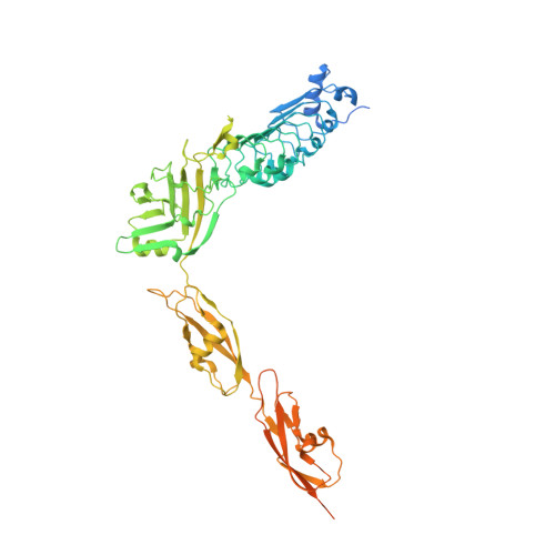
    Listeria monocytogenes is a human pathogen that employs a wide variety of virulence factors in order to adhere to, invade, and replicate within target cells. Internalins play key roles in processes ranging from adhesion to receptor recognition and are thus essential for infection. Recently, InlK, a surface-associated internalin, was shown to be involved in Listeria's ability to escape from autophagy by recruitment of the major vault protein (MVP) to the bacterial surface. Here, we report the structure of InlK, which harbors four domains arranged in the shape of a "bent arm". The structure supports a role for the "elbow" of InlK in partner recognition, as well as of two Ig-like pedestals intercalated by hinge regions in the projection of InlK away from the surface of the bacterium. The unusual fold and flexibility of InlK could be essential for MVP binding and concealment from recognition by molecules involved in the autophagic process.


  • Organizational Affiliation
    • Brazilian National Laboratory for Biosciences (LNBio), CNPEM, Campinas, São Paulo, Brazil.

Macromolecule Content 

  • Total Structure Weight: 129.44 kDa 
  • Atom Count: 6,832 
  • Modeled Residue Count: 864 
  • Deposited Residue Count: 1,180 
  • Unique protein chains: 1

Macromolecules

Find similar proteins by:|  3D Structure
Entity ID: 1
MoleculeChains  Sequence LengthOrganismDetailsImage
Internalin K
A, B
590Listeria monocytogenes EGD-eMutation(s): 0 
Gene Names: lmo1290
UniProt
Find proteins for Q8Y7I7 (Listeria monocytogenes serovar 1/2a (strain ATCC BAA-679 / EGD-e))
Explore Q8Y7I7 
Go to UniProtKB:  Q8Y7I7
Entity Groups
Sequence Clusters30% Identity50% Identity70% Identity90% Identity95% Identity100% Identity
UniProt GroupQ8Y7I7
Sequence Annotations
Expand
Reference Sequence

Small Molecules

Ligands 4 Unique
IDChains Name / Formula / InChI Key2D Diagram3D Interactions
GOL

Query on GOL


Download:Ideal Coordinates CCD File
G [auth B]GLYCEROL
C3 H8 O3
PEDCQBHIVMGVHV-UHFFFAOYSA-N
K

Query on K


Download:Ideal Coordinates CCD File
H [auth B]POTASSIUM ION
K
NPYPAHLBTDXSSS-UHFFFAOYSA-N
MG

Query on MG


Download:Ideal Coordinates CCD File
C [auth A],
D [auth A],
F [auth B]
MAGNESIUM ION
Mg
JLVVSXFLKOJNIY-UHFFFAOYSA-N
NA

Query on NA


Download:Ideal Coordinates CCD File
E [auth A]SODIUM ION
Na
FKNQFGJONOIPTF-UHFFFAOYSA-N

Experimental Data & Validation

Experimental Data

  • Method: X-RAY DIFFRACTION
  • Resolution: 2.59 Å
  • R-Value Free:  0.243 (Depositor), 0.244 (DCC) 
  • R-Value Work:  0.228 (Depositor), 0.230 (DCC) 
  • R-Value Observed: 0.230 (Depositor) 
Space Group: P 32 1 2
Unit Cell:
Length ( Å )Angle ( ˚ )
a = 115.34α = 90
b = 115.34β = 90
c = 190.71γ = 120
Software Package:
Software NamePurpose
ADSCdata collection
PHASERphasing
PHENIXrefinement
XDSdata reduction
XDSdata scaling

Structure Validation

View Full Validation Report


Entry History 

Deposition Data

  • Released Date: 2013-08-28 
  • Deposition Author(s): Neves, D.

Revision History  (Full details and data files)

  • Version 1.0: 2013-08-28
    Type: Initial release
  • Version 1.1: 2013-09-04
    Changes: Database references
  • Version 1.2: 2013-11-13
    Changes: Database references
  • Version 1.3: 2024-02-28
    Changes: Data collection, Database references, Derived calculations