4KXY | pdb_00004kxy

Human transketolase in complex with ThDP analogue (R)-2-(1,2-dihydroxyethyl)-3-deaza-ThDP


Experimental Data Snapshot

  • Method: X-RAY DIFFRACTION
  • Resolution: 1.26 Å
  • R-Value Free: 
    0.204 (Depositor), 0.182 (DCC) 
  • R-Value Work: 
    0.154 (Depositor), 0.149 (DCC) 
  • R-Value Observed: 
    0.154 (Depositor) 

Starting Model: experimental
View more details

wwPDB Validation 3D Report Full Report

Validation slider image for 4KXY

Ligand Structure Quality Assessment 


This is version 1.4 of the entry. See complete history

Literature

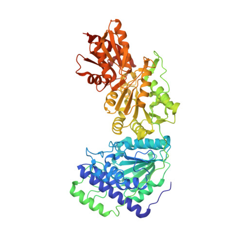
Sub-angstrom-resolution crystallography reveals physical distortions that enhance reactivity of a covalent enzymatic intermediate.

Ludtke, S.Neumann, P.Erixon, K.M.Leeper, F.Kluger, R.Ficner, R.Tittmann, K.

(2013) Nat Chem 5: 762-767

  • DOI: https://doi.org/10.1038/nchem.1728
  • Primary Citation Related Structures: 
    4KXU, 4KXV, 4KXW, 4KXX, 4KXY

  • PubMed Abstract: 

    It is recognized widely that enzymes promote reactions by providing a pathway that proceeds through a transition state of lower energy. In principle, further rate enhancements could be achieved if intermediates are prevented from relaxing to their lowest energy state, and thereby reduce the barrier to the subsequent transition state. Here, we report sub-ångström-resolution crystal structures of genuine covalent reaction intermediates of transketolase. These structures reveal a pronounced out-of-plane distortion of over 20° for the covalent bond that links cofactor and substrate, and a specific elongation of the scissile substrate carbon-carbon bond (d > 1.6 Å). To achieve these distortions, the protein's conformation appears to prevent relaxation of a substrate-cofactor intermediate. The results implicate a reduced barrier to the subsequent step that is consistent with an intermediate of raised energy and leads to a more efficient overall process.


  • Organizational Affiliation
    • Albrecht-von-Haller Institute, Göttingen Center for Molecular Biosciences, Georg-August University Göttingen, 37077 Göttingen, Germany.

Macromolecule Content 

  • Total Structure Weight: 142.26 kDa 
  • Atom Count: 11,716 
  • Modeled Residue Count: 1,240 
  • Deposited Residue Count: 1,274 
  • Unique protein chains: 1

Macromolecules

Find similar proteins by:|  3D Structure
Entity ID: 1
MoleculeChains  Sequence LengthOrganismDetailsImage
Transketolase
A, B
637Homo sapiensMutation(s): 0 
Gene Names: TKT
EC: 2.2.1.1
UniProt & NIH Common Fund Data Resources
Find proteins for P29401 (Homo sapiens)
Explore P29401 
Go to UniProtKB:  P29401
PHAROS:  P29401
GTEx:  ENSG00000163931 
Entity Groups
Sequence Clusters30% Identity50% Identity70% Identity90% Identity95% Identity100% Identity
UniProt GroupP29401
Sequence Annotations
Expand
Reference Sequence

Small Molecules

Ligands 5 Unique
IDChains Name / Formula / InChI Key2D Diagram3D Interactions
1U0

Query on 1U0


Download:Ideal Coordinates CCD File
K [auth A],
Z [auth B]
2-{4-[(4-amino-2-methylpyrimidin-5-yl)methyl]-5-[(1R)-1,2-dihydroxyethyl]-3-methylthiophen-2-yl}ethyl trihydrogen diphosphate
C15 H23 N3 O9 P2 S
AZNRIYLXMXTRTE-GFCCVEGCSA-N
SO4

Query on SO4


Download:Ideal Coordinates CCD File
GA [auth B],
R [auth A]
SULFATE ION
O4 S
QAOWNCQODCNURD-UHFFFAOYSA-L
EDO

Query on EDO


Download:Ideal Coordinates CCD File
BA [auth B]
C [auth A]
CA [auth B]
D [auth A]
DA [auth B]
BA [auth B],
C [auth A],
CA [auth B],
D [auth A],
DA [auth B],
E [auth A],
EA [auth B],
F [auth A],
FA [auth B],
G [auth A],
H [auth A],
IA [auth B],
JA [auth B],
KA [auth B],
L [auth A],
M [auth A],
N [auth A],
O [auth A],
P [auth A],
Q [auth A],
S [auth A],
T [auth A],
U [auth B],
V [auth B],
W [auth B],
X [auth B],
Y [auth B]
1,2-ETHANEDIOL
C2 H6 O2
LYCAIKOWRPUZTN-UHFFFAOYSA-N
CA

Query on CA


Download:Ideal Coordinates CCD File
AA [auth B],
I [auth A]
CALCIUM ION
Ca
BHPQYMZQTOCNFJ-UHFFFAOYSA-N
NA

Query on NA


Download:Ideal Coordinates CCD File
HA [auth B],
J [auth A]
SODIUM ION
Na
FKNQFGJONOIPTF-UHFFFAOYSA-N

Experimental Data & Validation

Experimental Data

  • Method: X-RAY DIFFRACTION
  • Resolution: 1.26 Å
  • R-Value Free:  0.204 (Depositor), 0.182 (DCC) 
  • R-Value Work:  0.154 (Depositor), 0.149 (DCC) 
  • R-Value Observed: 0.154 (Depositor) 
Space Group: P 1 21 1
Unit Cell:
Length ( Å )Angle ( ˚ )
a = 73.15α = 90
b = 86.05β = 94.12
c = 92.73γ = 90
Software Package:
Software NamePurpose
XSCALEdata scaling
SHELXrefinement
PDB_EXTRACTdata extraction
DNAdata collection
XDSdata reduction
PHENIXphasing
SHELXLrefinement

Structure Validation

View Full Validation Report


Ligand Structure Quality Assessment 


Entry History 

Deposition Data

Revision History  (Full details and data files)

  • Version 1.0: 2013-08-21
    Type: Initial release
  • Version 1.1: 2013-09-04
    Changes: Database references
  • Version 1.2: 2017-11-15
    Changes: Refinement description
  • Version 1.3: 2019-09-04
    Changes: Data collection
  • Version 1.4: 2023-09-20
    Changes: Data collection, Database references, Derived calculations, Refinement description