4KVN | pdb_00004kvn

Crystal structure of Fab 39.29 in complex with Influenza Hemagglutinin A/Perth/16/2009 (H3N2)


Experimental Data Snapshot

  • Method: X-RAY DIFFRACTION
  • Resolution: 3.10 Å
  • R-Value Free: 
    0.248 (Depositor), 0.253 (DCC) 
  • R-Value Work: 
    0.202 (Depositor), 0.214 (DCC) 
  • R-Value Observed: 
    0.205 (Depositor) 

Starting Model: experimental
View more details

wwPDB Validation 3D Report Full Report

Validation slider image for 4KVN

Ligand Structure Quality Assessment 


This is version 2.2 of the entry. See complete history

Literature

A Novel In vivo Human Plasmablast Enrichment Technique Allows Rapid Identification of Therapeutic Anti-Influenza A Antibodies

Nakamura, G.Chai, N.Park, S.Chiang, N.Lin, Z.Chiu, H.Fong, R.Yan, D.Kim, J.Zhang, J.Lee, W.P.Estevez, A.Coons, M.Xu, M.Lupardus, P.Balazs, M.Swem, L.R.

(2013) Cell Host Microbe 14: 93-103

  • DOI: https://doi.org/10.1016/j.chom.2013.06.004
  • Primary Citation Related Structures: 
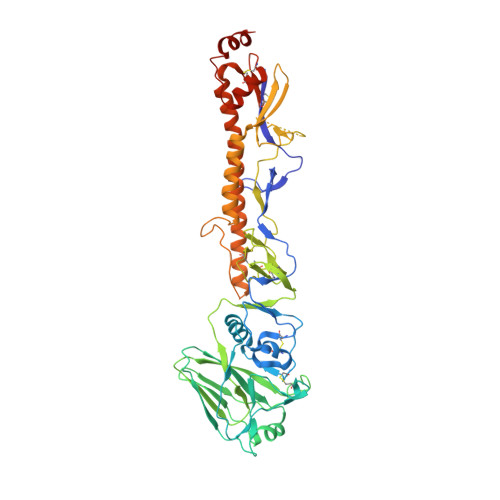
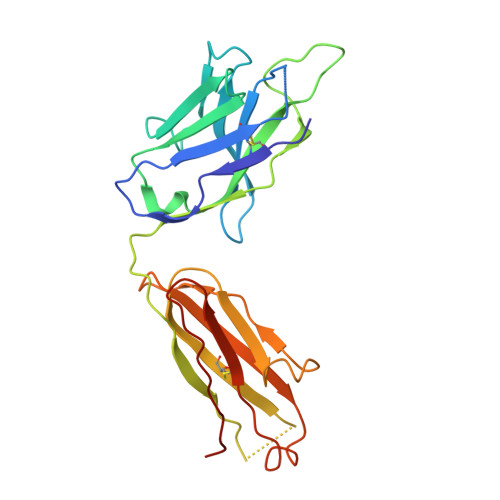
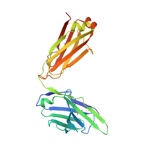
    4KVN

  • PubMed Abstract: 

    Recent advances enabling the cloning of human immunoglobulin G genes have proven effective for discovering monoclonal antibodies with therapeutic potential. However, these antibody-discovery methods are often arduous and identify only a few candidates from numerous antibody-secreting plasma cells or plasmablasts. We describe an in vivo enrichment technique that identifies broadly neutralizing human antibodies with high frequency. For this technique, human peripheral blood mononuclear cells from vaccinated donors are activated and enriched in an antigen-specific manner for the production of numerous antigen-specific plasmablasts. Using this technology, we identified four broadly neutralizing influenza A antibodies by screening only 840 human antibodies. Two of these antibodies neutralize every influenza A human isolate tested and perform better than the current anti-influenza A therapeutic, oseltamivir, in treating severe influenza infection in mice and ferrets. Furthermore, these antibodies elicit robust in vivo synergism when combined with oseltamivir, thus highlighting treatment strategies that could benefit influenza-infected patients.


  • Organizational Affiliation
    • Antibody Engineering Department, Genentech, South San Francisco, CA 94080, USA.

Macromolecule Content 

  • Total Structure Weight: 107.19 kDa 
  • Atom Count: 7,147 
  • Modeled Residue Count: 899 
  • Deposited Residue Count: 947 
  • Unique protein chains: 3

Macromolecules

Find similar proteins by:|  3D Structure
Entity ID: 1
MoleculeChains  Sequence LengthOrganismDetailsImage
Hemagglutinin505Influenza A virus (A/Perth/16/2009(H3N2))Mutation(s): 0 
Gene Names: HAHemagglutinin
UniProt
Find proteins for C6KNH7 (Influenza A virus)
Explore C6KNH7 
Go to UniProtKB:  C6KNH7
Entity Groups
Sequence Clusters30% Identity50% Identity70% Identity90% Identity95% Identity100% Identity
UniProt GroupC6KNH7
Glycosylation
Glycosylation Sites: 8
Sequence Annotations
Expand
Reference Sequence
Find similar proteins by:|  3D Structure
Entity ID: 2
MoleculeChains  Sequence LengthOrganismDetailsImage
Human IgG Heavy ChainB [auth H]227Homo sapiensMutation(s): 0 
Find similar proteins by:|  3D Structure
Entity ID: 3
MoleculeChains  Sequence LengthOrganismDetailsImage
Human IgG Light ChainC [auth L]215Homo sapiensMutation(s): 0 

Oligosaccharides

Help  
Entity ID: 4
MoleculeChains Length2D Diagram GlycosylationD Interactions
beta-D-mannopyranose-(1-4)-2-acetamido-2-deoxy-beta-D-glucopyranose-(1-4)-2-acetamido-2-deoxy-beta-D-glucopyranoseD [auth B],
E [auth C]
3N-Glycosylation
Glycosylation Resources
GlyTouCan: G15407YE
GlyCosmos: G15407YE
GlyGen: G15407YE
Entity ID: 5
MoleculeChains Length2D Diagram GlycosylationD Interactions
2-acetamido-2-deoxy-beta-D-glucopyranose-(1-4)-2-acetamido-2-deoxy-beta-D-glucopyranoseF [auth D]2N-Glycosylation
Glycosylation Resources
GlyTouCan: G42666HT
GlyCosmos: G42666HT
GlyGen: G42666HT

Small Molecules

Ligands 3 Unique
IDChains Name / Formula / InChI Key2D Diagram3D Interactions
NAG

Query on NAG


Download:Ideal Coordinates CCD File
G [auth A],
H [auth A],
I [auth A],
J [auth A],
K [auth A]
2-acetamido-2-deoxy-beta-D-glucopyranose
C8 H15 N O6
OVRNDRQMDRJTHS-FMDGEEDCSA-N
PGE

Query on PGE


Download:Ideal Coordinates CCD File
M [auth L]TRIETHYLENE GLYCOL
C6 H14 O4
ZIBGPFATKBEMQZ-UHFFFAOYSA-N
PO4

Query on PO4


Download:Ideal Coordinates CCD File
L
PHOSPHATE ION
O4 P
NBIIXXVUZAFLBC-UHFFFAOYSA-K

Experimental Data & Validation

Experimental Data

  • Method: X-RAY DIFFRACTION
  • Resolution: 3.10 Å
  • R-Value Free:  0.248 (Depositor), 0.253 (DCC) 
  • R-Value Work:  0.202 (Depositor), 0.214 (DCC) 
  • R-Value Observed: 0.205 (Depositor) 
Space Group: I 21 3
Unit Cell:
Length ( Å )Angle ( ˚ )
a = 204.37α = 90
b = 204.37β = 90
c = 204.37γ = 90
Software Package:
Software NamePurpose
PHASERphasing
BUSTERrefinement
MOSFLMdata reduction
SCALAdata scaling

Structure Validation

View Full Validation Report


Ligand Structure Quality Assessment 


Entry History 

Deposition Data

Revision History  (Full details and data files)

  • Version 1.0: 2013-08-07
    Type: Initial release
  • Version 1.1: 2020-03-04
    Changes: Advisory, Data collection, Database references, Derived calculations
  • Version 2.0: 2020-07-29
    Type: Remediation
    Reason: Carbohydrate remediation
    Changes: Advisory, Atomic model, Data collection, Derived calculations, Structure summary
  • Version 2.1: 2023-09-20
    Changes: Advisory, Data collection, Database references, Refinement description, Structure summary
  • Version 2.2: 2024-11-20
    Changes: Structure summary