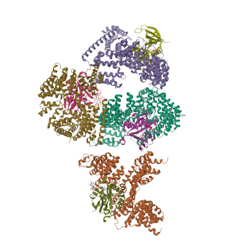
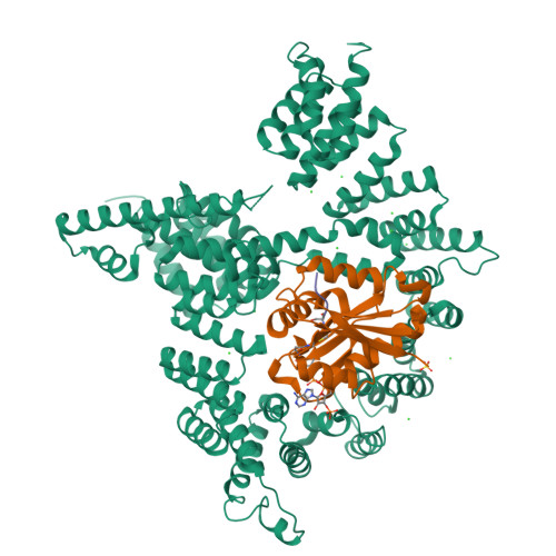
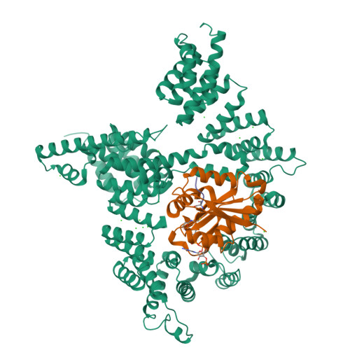
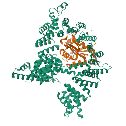
4KVM | pdb_00004kvm

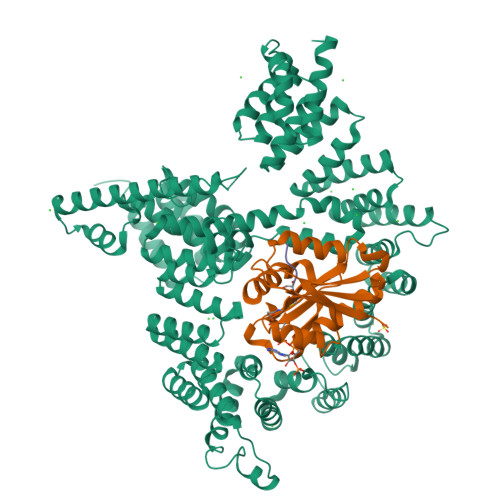
The NatA (Naa10p/Naa15p) amino-terminal acetyltransferase complex bound to a bisubstrate analog


Experimental Data Snapshot

  • Method: X-RAY DIFFRACTION
  • Resolution: 2.60 Å
  • R-Value Free: 
    0.260 (Depositor), 0.250 (DCC) 
  • R-Value Work: 
    0.222 (Depositor), 0.210 (DCC) 
  • R-Value Observed: 
    0.224 (Depositor) 

Starting Model: experimental
View more details

wwPDB Validation   3D Report Full Report


Ligand Structure Quality Assessment 

Created with Raphaël 2.3.0Worse 01 BetterLigand structure goodness of fit to experimental dataBest fitted 1XEClick on this verticalbar to view details

This is version 2.2 of the entry. See complete history


Literature

Molecular basis for N-terminal acetylation by the heterodimeric NatA complex.

Liszczak, G.Goldberg, J.M.Foyn, H.Petersson, E.J.Arnesen, T.Marmorstein, R.

(2013) Nat Struct Mol Biol 20: 1098-1105

  • DOI: https://doi.org/10.1038/nsmb.2636
  • Primary Citation of Related Structures:  
    4KVM, 4KVO, 4KVX

  • PubMed Abstract: 

    N-terminal acetylation is ubiquitous among eukaryotic proteins and controls a myriad of biological processes. Of the N-terminal acetyltransferases (NATs) that facilitate this cotranslational modification, the heterodimeric NatA complex has the most diversity for substrate selection and modifies the majority of all N-terminally acetylated proteins. Here, we report the X-ray crystal structure of the 100-kDa holo-NatA complex from Schizosaccharomyces pombe, in the absence and presence of a bisubstrate peptide-CoA-conjugate inhibitor, as well as the structure of the uncomplexed Naa10p catalytic subunit. The NatA-Naa15p auxiliary subunit contains 13 tetratricopeptide motifs and adopts a ring-like topology that wraps around the NatA-Naa10p subunit, an interaction that alters the Naa10p active site for substrate-specific acetylation. These studies have implications for understanding the mechanistic details of other NAT complexes and how regulatory subunits modulate the activity of the broader family of protein acetyltransferases.


  • Organizational Affiliation

    1] Program in Gene Expression and Regulation, Wistar Institute, Philadelphia, Pennsylvania, USA. [2] Department of Chemistry, University of Pennsylvania, Philadelphia, Pennsylvania, USA.


Macromolecules
Find similar proteins by:  (by identity cutoff)  |  3D Structure
Entity ID: 1
MoleculeChains Sequence LengthOrganismDetailsImage
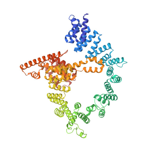
N-terminal acetyltransferase A complex subunit nat1
A, B, C, D
734Schizosaccharomyces pombe 972h-Mutation(s): 0 
Gene Names: nat1SPCC338.07c
UniProt
Find proteins for O74985 (Schizosaccharomyces pombe (strain 972 / ATCC 24843))
Explore O74985 
Go to UniProtKB:  O74985
Entity Groups  
Sequence Clusters30% Identity50% Identity70% Identity90% Identity95% Identity100% Identity
UniProt GroupO74985
Sequence Annotations
Expand
  • Reference Sequence
Find similar proteins by:  (by identity cutoff)  |  3D Structure
Entity ID: 2
MoleculeChains Sequence LengthOrganismDetailsImage
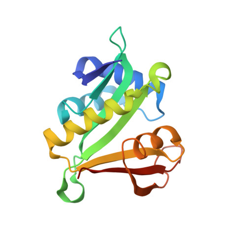
N-terminal acetyltransferase A complex catalytic subunit ard1
E, F, G, H
156Schizosaccharomyces pombe 972h-Mutation(s): 0 
Gene Names: ard1SPAC15E1.08
EC: 2.3.1.255
UniProt
Find proteins for Q9UTI3 (Schizosaccharomyces pombe (strain 972 / ATCC 24843))
Explore Q9UTI3 
Go to UniProtKB:  Q9UTI3
Entity Groups  
Sequence Clusters30% Identity50% Identity70% Identity90% Identity95% Identity100% Identity
UniProt GroupQ9UTI3
Sequence Annotations
Expand
  • Reference Sequence

Find similar proteins by:  Sequence   |   3D Structure  

Entity ID: 3
MoleculeChains Sequence LengthOrganismDetailsImage
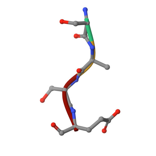
bisubstrate analog inhibitor
I, J, K, L
4synthetic constructMutation(s): 0 
Sequence Annotations
Expand
  • Reference Sequence
Small Molecules
Ligands 3 Unique
IDChains Name / Formula / InChI Key2D Diagram3D Interactions
1XE
Query on 1XE

Download Ideal Coordinates CCD File 
AC [auth I],
BC [auth J],
CC [auth K],
DC [auth L]
[5-(6-amino-9H-purin-9-yl)-4-hydroxy-3-(phosphonooxy)furan-2-yl]methyl (3R)-4-{[3-({(E)-2-[(2,2-dihydroxyethyl)sulfanyl]ethenyl}amino)-3-oxopropyl]amino}-3-hydroxy-2,2-dimethyl-4-oxobutyl dihydrogen diphosphate
C23 H34 N7 O18 P3 S
IWZWXZGRAJOFTE-QWNKOJSDSA-N
SO4
Query on SO4

Download Ideal Coordinates CCD File 
AA [auth A],
TB [auth D]
SULFATE ION
O4 S
QAOWNCQODCNURD-UHFFFAOYSA-L
CL
Query on CL

Download Ideal Coordinates CCD File 
AB [auth C]
BA [auth B]
BB [auth C]
CA [auth B]
CB [auth C]
CHLORIDE ION
Cl
VEXZGXHMUGYJMC-UHFFFAOYSA-M
Experimental Data & Validation

Experimental Data

  • Method: X-RAY DIFFRACTION
  • Resolution: 2.60 Å
  • R-Value Free:  0.260 (Depositor), 0.250 (DCC) 
  • R-Value Work:  0.222 (Depositor), 0.210 (DCC) 
  • R-Value Observed: 0.224 (Depositor) 
Space Group: P 1
Unit Cell:
Length ( Å )Angle ( ˚ )
a = 81.439α = 80.2
b = 119.381β = 76.6
c = 134.063γ = 70.42
Software Package:
Software NamePurpose
HKL-2000data collection
PHASERphasing
PHENIXrefinement
HKL-2000data reduction
HKL-2000data scaling

Structure Validation

View Full Validation Report


Ligand Structure Quality Assessment 

Created with Raphaël 2.3.0Worse 01 BetterLigand structure goodness of fit to experimental dataBest fitted 1XEClick on this verticalbar to view details

Entry History 

Deposition Data

Revision History  (Full details and data files)

  • Version 1.0: 2013-07-31
    Type: Initial release
  • Version 1.1: 2013-08-28
    Changes: Database references
  • Version 1.2: 2013-10-23
    Changes: Database references
  • Version 2.0: 2021-09-22
    Changes: Advisory, Database references, Derived calculations, Non-polymer description, Polymer sequence, Source and taxonomy, Structure summary
  • Version 2.1: 2023-09-20
    Changes: Data collection, Refinement description
  • Version 2.2: 2024-10-30
    Changes: Structure summary