4KSI | pdb_00004ksi

Crystal Structure Analysis of the Acidic Leucine Aminopeptidase of Tomato


Experimental Data Snapshot

  • Method: X-RAY DIFFRACTION
  • Resolution: 2.20 Å
  • R-Value Free: 
    0.173 (Depositor), 0.173 (DCC) 
  • R-Value Work: 
    0.137 (Depositor), 0.137 (DCC) 
  • R-Value Observed: 
    0.139 (Depositor) 

wwPDB Validation 3D Report Full Report

Validation slider image for 4KSI

This is version 1.4 of the entry. See complete history

Literature

Structure of tomato wound-induced leucine aminopeptidase sheds light on substrate specificity.

Duprez, K.Scranton, M.A.Walling, L.L.Fan, L.

(2014) Acta Crystallogr D Biol Crystallogr 70: 1649-1658

  • DOI: https://doi.org/10.1107/S1399004714006245
  • Primary Citation Related Structures: 
    4KSI

  • PubMed Abstract: 

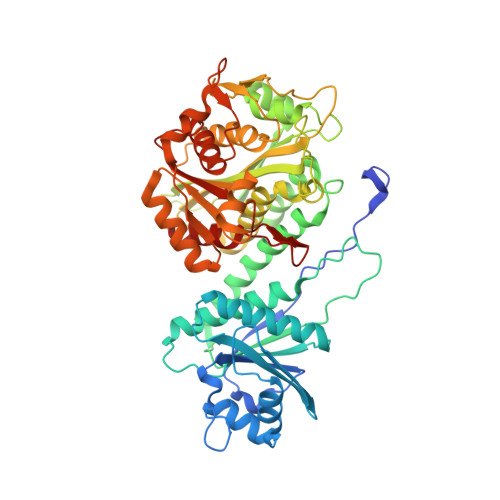
    The acidic leucine aminopeptidase (LAP-A) from tomato is induced in response to wounding and insect feeding. Although LAP-A shows in vitro peptidase activity towards peptides and peptide analogs, it is not clear what kind of substrates LAP-A hydrolyzes in vivo. In the current study, the crystal structure of LAP-A was determined to 2.20 Å resolution. Like other LAPs in the M17 peptidase family, LAP-A is a dimer of trimers containing six monomers of bilobal structure. Each monomer contains two metal ions bridged by a water or a hydroxyl ion at the active site. Modeling of different peptides or peptide analogs in the active site of LAP-A reveals a spacious substrate-binding channel that can bind peptides of five or fewer residues with few geometric restrictions. The sequence specificity of the bound peptide is likely to be selected by the structural and chemical restrictions on the amino acid at the P1 and P1' positions because these two amino acids have to bind perfectly at the active site for hydrolysis of the first peptide bond to occur. The hexameric assembly results in the merger of the open ends of the six substrate-binding channels from the LAP-A monomers to form a spacious central cavity allowing the hexameric LAP-A enzyme to simultaneously hydrolyze six peptides containing up to six amino acids each. The hexameric LAP-A enzyme may also hydrolyze long peptides or proteins if only one such substrate is bound to the hexamer because the substrate can extend through the central cavity and the two major solvent channels between the two LAP-A trimers.


  • Organizational Affiliation
    • Department of Biochemistry, University of California-Riverside, Riverside, CA 92521, USA.

Macromolecule Content 

  • Total Structure Weight: 56.79 kDa 
  • Atom Count: 4,567 
  • Modeled Residue Count: 517 
  • Deposited Residue Count: 529 
  • Unique protein chains: 1

Macromolecules

Find similar proteins by:|  3D Structure
Entity ID: 1
MoleculeChains  Sequence LengthOrganismDetailsImage
Leucine aminopeptidase 1, chloroplastic529Solanum lycopersicumMutation(s): 0 
Gene Names: LAPLAP2LAPA1
EC: 3.4.11.1 (PDB Primary Data), 3.4.11.5 (PDB Primary Data)
UniProt
Find proteins for Q10712 (Solanum lycopersicum)
Explore Q10712 
Go to UniProtKB:  Q10712
Entity Groups
Sequence Clusters30% Identity50% Identity70% Identity90% Identity95% Identity100% Identity
UniProt GroupQ10712
Sequence Annotations
Expand
Reference Sequence

Small Molecules

Ligands 6 Unique
IDChains Name / Formula / InChI Key2D Diagram3D Interactions
MPD

Query on MPD


Download:Ideal Coordinates CCD File
N [auth A](4S)-2-METHYL-2,4-PENTANEDIOL
C6 H14 O2
SVTBMSDMJJWYQN-YFKPBYRVSA-N
SO4

Query on SO4


Download:Ideal Coordinates CCD File
D [auth A],
O [auth A]
SULFATE ION
O4 S
QAOWNCQODCNURD-UHFFFAOYSA-L
GOL

Query on GOL


Download:Ideal Coordinates CCD File
H [auth A],
I [auth A],
J [auth A],
K [auth A],
L [auth A]
GLYCEROL
C3 H8 O3
PEDCQBHIVMGVHV-UHFFFAOYSA-N
IMD

Query on IMD


Download:Ideal Coordinates CCD File
M [auth A]IMIDAZOLE
C3 H5 N2
RAXXELZNTBOGNW-UHFFFAOYSA-O
CL

Query on CL


Download:Ideal Coordinates CCD File
E [auth A],
F [auth A],
G [auth A]
CHLORIDE ION
Cl
VEXZGXHMUGYJMC-UHFFFAOYSA-M
MG

Query on MG


Download:Ideal Coordinates CCD File
B [auth A],
C [auth A]
MAGNESIUM ION
Mg
JLVVSXFLKOJNIY-UHFFFAOYSA-N

Experimental Data & Validation

Experimental Data

  • Method: X-RAY DIFFRACTION
  • Resolution: 2.20 Å
  • R-Value Free:  0.173 (Depositor), 0.173 (DCC) 
  • R-Value Work:  0.137 (Depositor), 0.137 (DCC) 
  • R-Value Observed: 0.139 (Depositor) 
Space Group: P 63 2 2
Unit Cell:
Length ( Å )Angle ( ˚ )
a = 160.597α = 90
b = 160.597β = 90
c = 104.183γ = 120
Software Package:
Software NamePurpose
CrystalCleardata collection
PHASERphasing
REFMACrefinement
HKL-2000data reduction
HKL-2000data scaling

Structure Validation

View Full Validation Report


Entry History 

Deposition Data

Revision History  (Full details and data files)

  • Version 1.0: 2013-06-12
    Type: Initial release
  • Version 1.1: 2014-06-18
    Changes: Database references
  • Version 1.2: 2014-09-24
    Changes: Database references
  • Version 1.3: 2017-11-15
    Changes: Refinement description
  • Version 1.4: 2024-02-28
    Changes: Data collection, Database references, Derived calculations