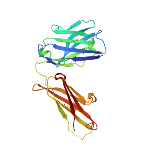
4KPH

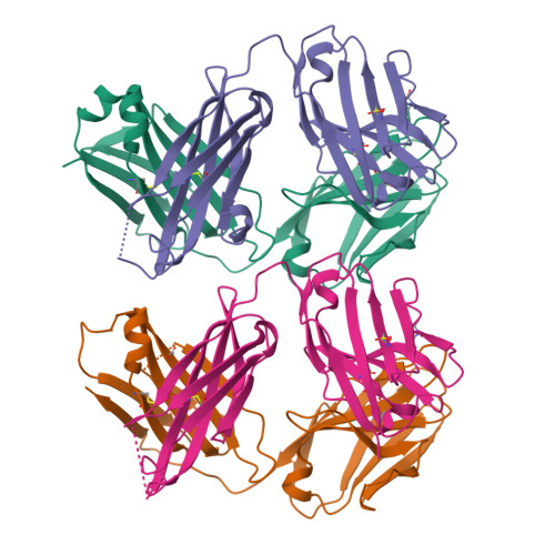
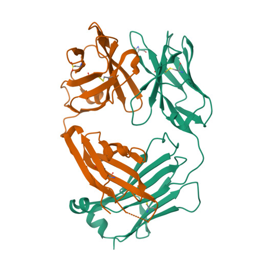
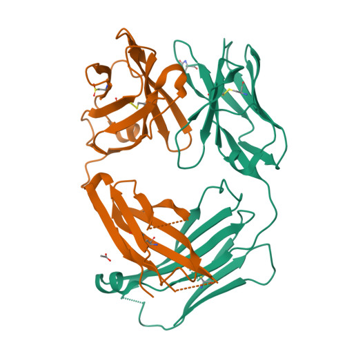
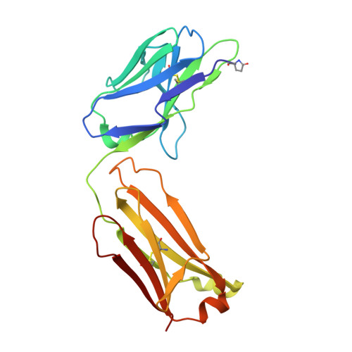
Structure of the Fab fragment of N62, a protective monoclonal antibody to the nonreducing end of Francisella tularensis O-antigen


Experimental Data Snapshot

  • Method: X-RAY DIFFRACTION
  • Resolution: 2.59 Å
  • R-Value Free: 0.268 
  • R-Value Work: 0.222 
  • R-Value Observed: 0.225 

Starting Models: experimental
View more details

wwPDB Validation   3D Report Full Report


This is version 2.1 of the entry. See complete history


Literature

The binding sites of monoclonal antibodies to the non-reducing end of Francisella tularensis O-antigen accommodate mainly the terminal saccharide.

Lu, Z.Rynkiewicz, M.J.Yang, C.Y.Madico, G.Perkins, H.M.Wang, Q.Costello, C.E.Zaia, J.Seaton, B.A.Sharon, J.

(2013) Immunology 140: 374-389

  • DOI: https://doi.org/10.1111/imm.12150
  • Primary Citation of Related Structures:  
    4KPH

  • PubMed Abstract: 

    We have previously described two types of protective B-cell epitopes in the O-antigen (OAg) of the Gram-negative bacterium Francisella tularensis: repeating internal epitopes targeted by the vast majority of anti-OAg monoclonal antibodies (mAbs), and a non-overlapping epitope at the non-reducing end targeted by the previously unique IgG2a mAb FB11. We have now generated and characterized three mAbs specific for the non-reducing end of F. tularensis OAg, partially encoded by the same variable region germline genes, indicating that they target the same epitope. Like FB11, the new mAbs, Ab63 (IgG3), N213 (IgG3) and N62 (IgG2b), had higher antigen-binding bivalent avidity than internally binding anti-OAg mAbs, and an oligosaccharide containing a single OAg repeat was sufficient for optimal inhibition of their antigen-binding. The X-ray crystal structure of N62 Fab showed that the antigen-binding site is lined mainly by aromatic amino acids that form a small cavity, which can accommodate no more than one and a third sugar residues, indicating that N62 binds mainly to the terminal Qui4NFm residue at the nonreducing end of OAg. In efficacy studies with mice infected intranasally with the highly virulent F. tularensis strain SchuS4, N62, N213 and Ab63 prolonged survival and reduced blood bacterial burden. These results yield insights into how antibodies to non-reducing ends of microbial polysaccharides can contribute to immune protection despite the smaller size of their target epitopes compared with antibodies to internal polysaccharide regions.


  • Organizational Affiliation

    Department of Pathology and Laboratory Medicine, Boston University School of Medicine, Boston, MA, USA.


Macromolecules
Find similar proteins by:  (by identity cutoff)  |  3D Structure
Entity ID: 1
MoleculeChains Sequence LengthOrganismDetailsImage
N62 light chainA [auth L],
C [auth M]
210Mus musculusMutation(s): 0 
Entity Groups  
Sequence Clusters30% Identity50% Identity70% Identity90% Identity95% Identity100% Identity
Sequence Annotations
Expand
  • Reference Sequence
Find similar proteins by:  (by identity cutoff)  |  3D Structure
Entity ID: 2
MoleculeChains Sequence LengthOrganismDetailsImage
N62 heavy chainB [auth H],
D [auth I]
212Mus musculusMutation(s): 0 
Entity Groups  
Sequence Clusters30% Identity50% Identity70% Identity90% Identity95% Identity100% Identity
Sequence Annotations
Expand
  • Reference Sequence
Small Molecules
Ligands 1 Unique
IDChains Name / Formula / InChI Key2D Diagram3D Interactions
ACT
Query on ACT

Download Ideal Coordinates CCD File 
E [auth M]ACETATE ION
C2 H3 O2
QTBSBXVTEAMEQO-UHFFFAOYSA-M
Modified Residues  2 Unique
IDChains TypeFormula2D DiagramParent
PCA
Query on PCA
A [auth L],
C [auth M]
L-PEPTIDE LINKINGC5 H7 N O3GLN
CSD
Query on CSD
B [auth H],
D [auth I]
L-PEPTIDE LINKINGC3 H7 N O4 SCYS
Experimental Data & Validation

Experimental Data

  • Method: X-RAY DIFFRACTION
  • Resolution: 2.59 Å
  • R-Value Free: 0.268 
  • R-Value Work: 0.222 
  • R-Value Observed: 0.225 
  • Space Group: P 21 21 21
Unit Cell:
Length ( Å )Angle ( ˚ )
a = 68.874α = 90
b = 87.076β = 90
c = 153.589γ = 90
Software Package:
Software NamePurpose
DENZOdata reduction
SCALEPACKdata scaling
PHASERphasing
PHENIXrefinement
PDB_EXTRACTdata extraction

Structure Validation

View Full Validation Report


Entry History 

Deposition Data

Revision History  (Full details and data files)

  • Version 1.0: 2013-07-24
    Type: Initial release
  • Version 1.1: 2013-10-23
    Changes: Database references
  • Version 2.0: 2019-12-25
    Changes: Derived calculations, Polymer sequence
  • Version 2.1: 2023-09-20
    Changes: Data collection, Database references, Derived calculations, Refinement description