4KMG

Crystal structure of cytochrome c6B from Synechococcus sp. WH8102


Experimental Data Snapshot

  • Method: X-RAY DIFFRACTION
  • Resolution: 1.40 Å
  • R-Value Free: 0.181 
  • R-Value Work: 0.120 
  • R-Value Observed: 0.124 

Starting Model: experimental
View more details

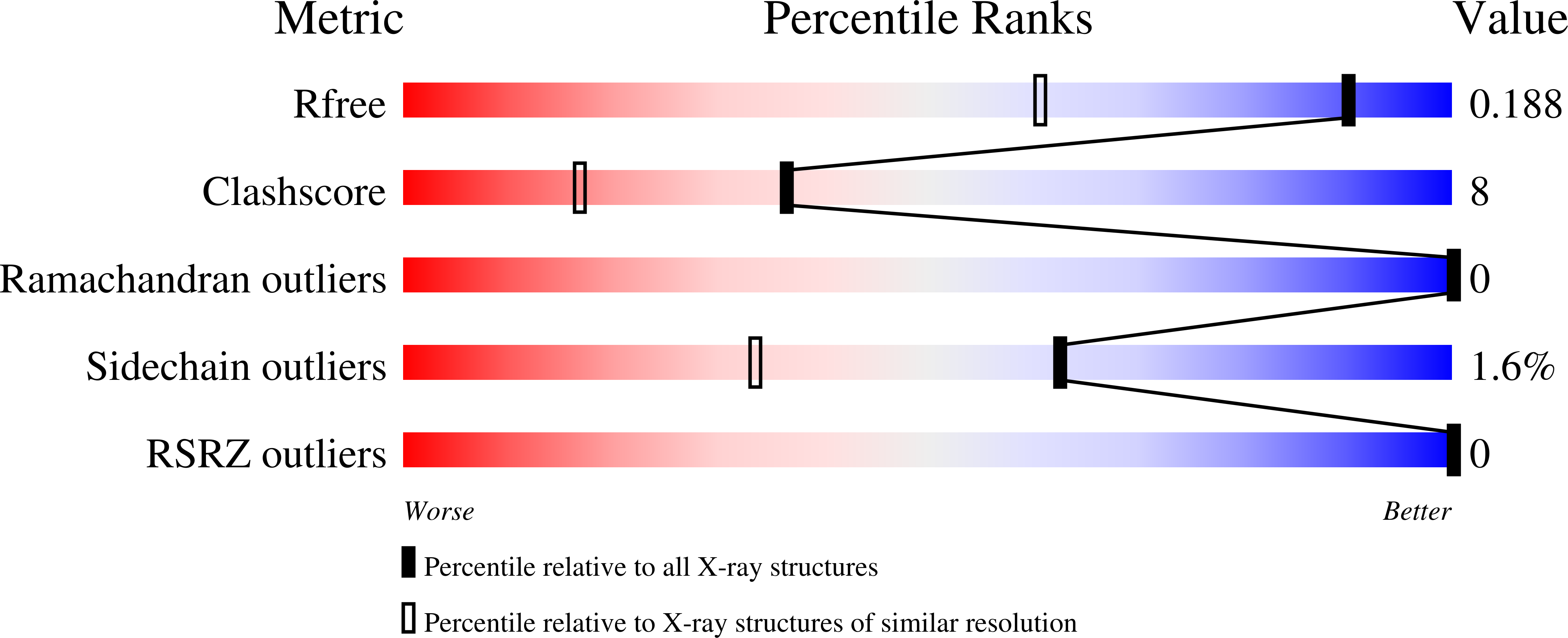
wwPDB Validation   3D Report Full Report


Ligand Structure Quality Assessment 


This is version 2.1 of the entry. See complete history


Literature

Cytochrome c6B of Synechococcus sp. WH 8102 - Crystal structure and basic properties of novel c6-like family representative.

Zatwarnicki, P.Barciszewski, J.Krzywda, S.Jaskolski, M.Kolesinski, P.Szczepaniak, A.

(2014) Biochem Biophys Res Commun 443: 1131-1135

  • DOI: https://doi.org/10.1016/j.bbrc.2013.10.167
  • Primary Citation of Related Structures:  
    4KMG

  • PubMed Abstract: 

    Cytochromes c are soluble electron carriers of relatively low molecular weight, containing single heme moiety. In cyanobacteria cytochrome c6 participates in electron transfer from cytochrome b6f complex to photosystem I. Recent phylogenetic analysis revealed the existence of a few families of proteins homologous to the previously mentioned. Cytochrome c6A from Arabidopsis thaliana was identified as a protein responsible for disulfide bond formation in response to intracellular redox state changes and c550 is well known element of photosystem II. However, function of cytochromes marked as c6B, c6C and cM as well as the physiological process in which they take a part still remain unidentified. Here we present the first structural and biophysical analysis of cytochrome from the c6B family from mesophilic cyanobacteria Synechococcus sp. WH 8102. Purified protein was crystallized and its structure was refined at 1.4 Å resolution. Overall architecture of this polypeptide resembles typical I-class cytochromes c. The main features, that distinguish described protein from cytochrome c6, are slightly red-shifted α band of UV-Vis spectrum as well as relatively low midpoint potential (113.2±2.2 mV). Although, physiological function of cytochrome c6B has yet to be determined its properties probably exclude the participation of this protein in electron trafficking between b6f complex and photosystem I.


  • Organizational Affiliation

    Department of Biophysics, Faculty of Biotechnology, University of Wroclaw, F. Joliot Curie 14a, 50-383 Wroclaw, Poland.


Macromolecules
Find similar proteins by:  (by identity cutoff)  |  3D Structure
Entity ID: 1
MoleculeChains Sequence LengthOrganismDetailsImage
Cytochrome C6 (Soluble cytochrome F) (Cytochrome c553)88Parasynechococcus marenigrum WH 8102Mutation(s): 0 
Gene Names: NP_897609petJSYNW1516
UniProt
Find proteins for Q7U624 (Parasynechococcus marenigrum (strain WH8102))
Explore Q7U624 
Go to UniProtKB:  Q7U624
Entity Groups  
Sequence Clusters30% Identity50% Identity70% Identity90% Identity95% Identity100% Identity
UniProt GroupQ7U624
Sequence Annotations
Expand
  • Reference Sequence
Small Molecules
Ligands 2 Unique
IDChains Name / Formula / InChI Key2D Diagram3D Interactions
HEC
Query on HEC

Download Ideal Coordinates CCD File 
B [auth A]HEME C
C34 H34 Fe N4 O4
HXQIYSLZKNYNMH-LJNAALQVSA-N
NA
Query on NA

Download Ideal Coordinates CCD File 
C [auth A]SODIUM ION
Na
FKNQFGJONOIPTF-UHFFFAOYSA-N
Experimental Data & Validation

Experimental Data

  • Method: X-RAY DIFFRACTION
  • Resolution: 1.40 Å
  • R-Value Free: 0.181 
  • R-Value Work: 0.120 
  • R-Value Observed: 0.124 
  • Space Group: C 1 2 1
Unit Cell:
Length ( Å )Angle ( ˚ )
a = 106.11α = 90
b = 28.98β = 92.3
c = 24.68γ = 90
Software Package:
Software NamePurpose
MAR345dtbdata collection
MOLREPphasing
REFMACrefinement
XDSdata reduction
XDSdata scaling

Structure Validation

View Full Validation Report


Ligand Structure Quality Assessment 


Entry History 

Deposition Data

Revision History  (Full details and data files)

  • Version 1.0: 2014-03-26
    Type: Initial release
  • Version 1.1: 2017-11-15
    Changes: Advisory, Refinement description
  • Version 2.0: 2021-03-10
    Changes: Atomic model, Data collection, Derived calculations, Non-polymer description, Structure summary
  • Version 2.1: 2023-09-20
    Changes: Advisory, Data collection, Database references, Refinement description