4KM5 | pdb_00004km5

X-ray crystal structure of human cyclic GMP-AMP synthase (cGAS)


Experimental Data Snapshot

  • Method: X-RAY DIFFRACTION
  • Resolution: 2.50 Å
  • R-Value Free: 
    0.240 (Depositor), 0.240 (DCC) 
  • R-Value Work: 
    0.225 (Depositor), 0.229 (DCC) 
  • R-Value Observed: 
    0.225 (Depositor) 

wwPDB Validation 3D Report Full Report

Validation slider image for 4KM5

This is version 1.3 of the entry. See complete history

Literature

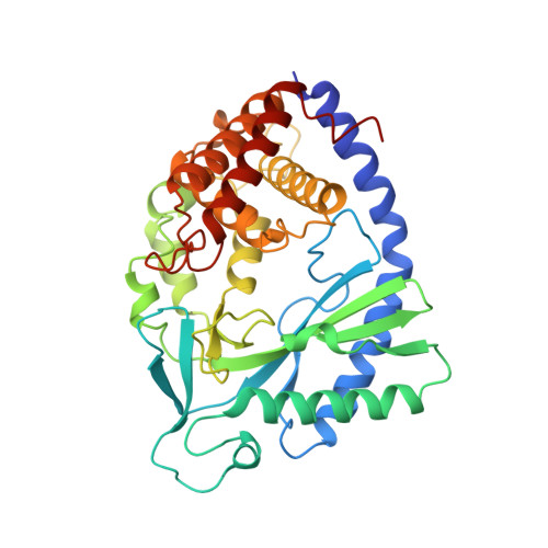
Structure of Human cGAS Reveals a Conserved Family of Second-Messenger Enzymes in Innate Immunity.

Kranzusch, P.J.Lee, A.S.Berger, J.M.Doudna, J.A.

(2013) Cell Rep 3: 1362-1368

  • DOI: https://doi.org/10.1016/j.celrep.2013.05.008
  • Primary Citation Related Structures: 
    4KM5

  • PubMed Abstract: 

    Innate immune recognition of foreign nucleic acids induces protective interferon responses. Detection of cytosolic DNA triggers downstream immune signaling through activation of cyclic GMP-AMP synthase (cGAS). We report here the crystal structure of human cGAS, revealing an unanticipated zinc-ribbon DNA-binding domain appended to a core enzymatic nucleotidyltransferase scaffold. The catalytic core of cGAS is structurally homologous to the RNA-sensing enzyme, 2'-5' oligo-adenylate synthase (OAS), and divergent C-terminal domains account for specific ligand-activation requirements of each enzyme. We show that the cGAS zinc ribbon is essential for STING-dependent induction of the interferon response and that conserved amino acids displayed within the intervening loops are required for efficient cytosolic DNA recognition. These results demonstrate that cGAS and OAS define a family of innate immunity sensors and that structural divergence from a core nucleotidyltransferase enables second-messenger responses to distinct foreign nucleic acids.


  • Organizational Affiliation
    • Department of Molecular & Cell Biology, Center for RNA Systems Biology, Howard Hughes Medical Institute, University of California, Berkeley, CA 94720, USA.

Macromolecule Content 

  • Total Structure Weight: 43.06 kDa 
  • Atom Count: 3,017 
  • Modeled Residue Count: 362 
  • Deposited Residue Count: 369 
  • Unique protein chains: 1

Macromolecules

Find similar proteins by:|  3D Structure
Entity ID: 1
MoleculeChains  Sequence LengthOrganismDetailsImage
Cyclic GMP-AMP synthase369Homo sapiensMutation(s): 0 
Gene Names: MB21D1C6orf150
EC: 2.7.7 (PDB Primary Data), 2.7.7.86 (UniProt)
UniProt & NIH Common Fund Data Resources
Find proteins for Q8N884 (Homo sapiens)
Explore Q8N884 
Go to UniProtKB:  Q8N884
PHAROS:  Q8N884
GTEx:  ENSG00000164430 
Entity Groups
Sequence Clusters30% Identity50% Identity70% Identity90% Identity95% Identity100% Identity
UniProt GroupQ8N884
Sequence Annotations
Expand
Reference Sequence

Small Molecules

Ligands 1 Unique
IDChains Name / Formula / InChI Key2D Diagram3D Interactions
ZN

Query on ZN


Download:Ideal Coordinates CCD File
B [auth A]ZINC ION
Zn
PTFCDOFLOPIGGS-UHFFFAOYSA-N

Experimental Data & Validation

Experimental Data

  • Method: X-RAY DIFFRACTION
  • Resolution: 2.50 Å
  • R-Value Free:  0.240 (Depositor), 0.240 (DCC) 
  • R-Value Work:  0.225 (Depositor), 0.229 (DCC) 
  • R-Value Observed: 0.225 (Depositor) 
Space Group: P 21 21 2
Unit Cell:
Length ( Å )Angle ( ˚ )
a = 46.454α = 90
b = 162.7β = 90
c = 58.407γ = 90
Software Package:
Software NamePurpose
Web-Icedata collection
PHENIXmodel building
PHENIXrefinement
XDSdata reduction
SCALAdata scaling
PHENIXphasing

Structure Validation

View Full Validation Report


Entry History 

Deposition Data

Revision History  (Full details and data files)

  • Version 1.0: 2013-05-29
    Type: Initial release
  • Version 1.1: 2013-06-12
    Changes: Database references
  • Version 1.2: 2017-11-15
    Changes: Refinement description
  • Version 1.3: 2024-02-28
    Changes: Data collection, Database references, Derived calculations