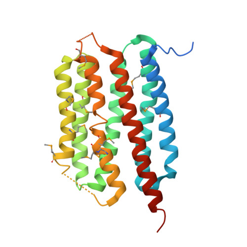
Cross-protomer interaction with the photoactive site in oligomeric proteorhodopsin complexes.
Ran, T., Ozorowski, G., Gao, Y., Sineshchekov, O.A., Wang, W., Spudich, J.L., Luecke, H.(2013) Acta Crystallogr D Biol Crystallogr 69: 1965-1980
- PubMed: 24100316
- DOI: https://doi.org/10.1107/S0907444913017575
- Primary Citation of Related Structures:
4JQ6, 4KLY, 4KNF - PubMed Abstract:
Proteorhodopsins (PRs), members of the microbial rhodopsin superfamily of seven-transmembrane-helix proteins that use retinal chromophores, comprise the largest subfamily of rhodopsins, yet very little structural information is available. PRs are ubiquitous throughout the biosphere and their genes have been sequenced in numerous species of bacteria. They have been shown to exhibit ion-pumping activity like their archaeal homolog bacteriorhodopsin (BR). Here, the first crystal structure of a proteorhodopsin, that of a blue-light-absorbing proteorhodopsin (BPR) isolated from the Mediterranean Sea at a depth of 12 m (Med12BPR), is reported. Six molecules of Med12BPR form a doughnut-shaped C6 hexameric ring, unlike BR, which forms a trimer. Furthermore, the structures of two mutants of a related BPR isolated from the Pacific Ocean near Hawaii at a depth of 75 m (HOT75BPR), which show a C5 pentameric arrangement, are reported. In all three structures the retinal polyene chain is shifted towards helix C when compared with other microbial rhodopsins, and the putative proton-release group in BPR differs significantly from those of BR and xanthorhodopsin (XR). The most striking feature of proteorhodopsin is the position of the conserved active-site histidine (His75, also found in XR), which forms a hydrogen bond to the proton acceptor from the same molecule (Asp97) and also to Trp34 of a neighboring protomer. Trp34 may function by stabilizing His75 in a conformation that favors a deprotonated Asp97 in the dark state, and suggests cooperative behavior between protomers when the protein is in an oligomeric form. Mutation-induced alterations in proton transfers in the BPR photocycle in Escherichia coli cells provide evidence for a similar cross-protomer interaction of BPR in living cells and a functional role of the inter-protomer Trp34-His75 interaction in ion transport. Finally, Wat402, a key molecule responsible for proton translocation between the Schiff base and the proton acceptor in BR, appears to be absent in PR, suggesting that the ion-transfer mechanism may differ between PR and BR.
- Key Laboratory of Agricultural and Environmental Microbiology, Ministry of Agriculture, College of Life Sciences, Nanjing Agricultural University, Nanjing 210095, People's Republic of China.
Organizational Affiliation:


