4KK4 | pdb_00004kk4

YwlE arginine phosphatase - C7S mutant with phosphorylated active site serine


Experimental Data Snapshot

  • Method: X-RAY DIFFRACTION
  • Resolution: 1.80 Å
  • R-Value Free: 
    0.215 (Depositor), 0.213 (DCC) 
  • R-Value Work: 
    0.172 (Depositor), 0.171 (DCC) 
  • R-Value Observed: 
    0.174 (Depositor) 

wwPDB Validation 3D Report Full Report

Validation slider image for 4KK4

This is version 1.2 of the entry. See complete history

Literature

Structural basis for recognizing phosphoarginine and evolving residue-specific protein phosphatases in gram-positive bacteria.

Fuhrmann, J.Mierzwa, B.Trentini, D.B.Spiess, S.Lehner, A.Charpentier, E.Clausen, T.

(2013) Cell Rep 3: 1832-1839

  • DOI: https://doi.org/10.1016/j.celrep.2013.05.023
  • Primary Citation Related Structures: 
    4KK3, 4KK4

  • PubMed Abstract: 

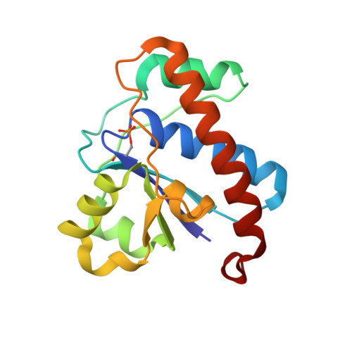
    Many cellular pathways are regulated by the competing activity of protein kinases and phosphatases. The recent identification of arginine phosphorylation as a protein modification in bacteria prompted us to analyze the molecular basis of targeting phospho-arginine. In this work, we characterize an annotated tyrosine phosphatase, YwlE, that counteracts the protein arginine kinase McsB. Strikingly, structural studies of YwlE reaction intermediates provide a direct view on a captured arginine residue. Together with biochemical data, the crystal structures depict the evolution of a highly specific phospho-arginine phosphatase, with the use of a size-and-polarity filter for distinguishing phosphorylated arginine from other phosphorylated side chains. To confirm the proposed mechanism, we performed bioinformatic searches for phosphatases, employing a similar selectivity filter, and identified a protein in Drosophila melanogaster exhibiting robust arginine phosphatase activity. In sum, our findings uncover the molecular framework for specific targeting of phospho-arginine and suggest that protein arginine (de)phosphorylation may be relevant in eukaryotes.


  • Organizational Affiliation
    • Research Institute of Molecular Pathology (IMP), A-1030 Vienna, Austria.

Macromolecule Content 

  • Total Structure Weight: 17.12 kDa 
  • Atom Count: 1,266 
  • Modeled Residue Count: 152 
  • Deposited Residue Count: 152 
  • Unique protein chains: 1

Macromolecules

Find similar proteins by:|  3D Structure
Entity ID: 1
MoleculeChains  Sequence LengthOrganismDetailsImage
Low molecular weight protein-tyrosine-phosphatase YwlE152Bacillus subtilis subsp. subtilis str. 168Mutation(s): 1 
Gene Names: ywlEBSU36930ipc-31d
EC: 3.1.3.48 (PDB Primary Data), 3.9.1.2 (UniProt)
UniProt
Find proteins for P39155 (Bacillus subtilis (strain 168))
Explore P39155 
Go to UniProtKB:  P39155
Entity Groups
Sequence Clusters30% Identity50% Identity70% Identity90% Identity95% Identity100% Identity
UniProt GroupP39155
Sequence Annotations
Expand
Reference Sequence

Small Molecules

Modified Residues  1 Unique
IDChains TypeFormula2D DiagramParent
SEP
Query on SEP
A
L-PEPTIDE LINKINGC3 H8 N O6 PSER

Experimental Data & Validation

Experimental Data

  • Method: X-RAY DIFFRACTION
  • Resolution: 1.80 Å
  • R-Value Free:  0.215 (Depositor), 0.213 (DCC) 
  • R-Value Work:  0.172 (Depositor), 0.171 (DCC) 
  • R-Value Observed: 0.174 (Depositor) 
Space Group: C 1 2 1
Unit Cell:
Length ( Å )Angle ( ˚ )
a = 85.84α = 90
b = 39.34β = 89.7
c = 40.67γ = 90
Software Package:
Software NamePurpose
DENZOdata reduction
SCALAdata scaling
PHENIXrefinement
PDB_EXTRACTdata extraction
DNAdata collection
PHASERphasing

Structure Validation

View Full Validation Report


Entry History 

Deposition Data

Revision History  (Full details and data files)

  • Version 1.0: 2013-07-03
    Type: Initial release
  • Version 1.1: 2013-07-17
    Changes: Database references
  • Version 1.2: 2024-11-20
    Changes: Data collection, Database references, Derived calculations, Structure summary