4KJI | pdb_00004kji

Novel re-arrangement of an RsmA/cSRa family protein to create a structurally distinct new RNA-binding family member


Experimental Data Snapshot

  • Method: X-RAY DIFFRACTION
  • Resolution: 3.20 Å
  • R-Value Free: 
    0.317 (Depositor), 0.324 (DCC) 
  • R-Value Work: 
    0.222 (Depositor), 0.230 (DCC) 
  • R-Value Observed: 
    0.226 (Depositor) 

Starting Models: experimental
View more details

wwPDB Validation 3D Report Full Report

Validation slider image for 4KJI

This is version 1.3 of the entry. See complete history

Literature

Structural Rearrangement in an RsmA/CsrA Ortholog of Pseudomonas aeruginosa Creates a Dimeric RNA-Binding Protein, RsmN.

Morris, E.R.Hall, G.Li, C.Heeb, S.Kulkarni, R.V.Lovelock, L.Silistre, H.Messina, M.Camara, M.Emsley, J.Williams, P.Searle, M.S.

(2013) Structure 21: 1659-1671

  • DOI: https://doi.org/10.1016/j.str.2013.07.007
  • Primary Citation Related Structures: 
    4KJI, 4KRW

  • PubMed Abstract: 

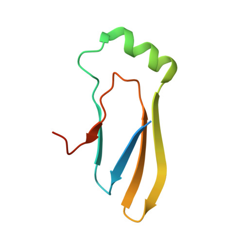
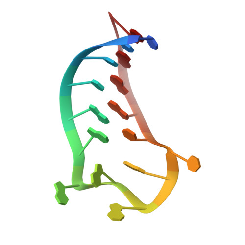
    In bacteria, the highly conserved RsmA/CsrA family of RNA-binding proteins functions as global posttranscriptional regulators acting on mRNA translation and stability. Through phenotypic complementation of an rsmA mutant in Pseudomonas aeruginosa, we discovered a family member, termed RsmN. Elucidation of the RsmN crystal structure and that of the complex with a hairpin from the sRNA, RsmZ, reveals a uniquely inserted α helix, which redirects the polypeptide chain to form a distinctly different protein fold to the domain-swapped dimeric structure of RsmA homologs. The overall β sheet structure required for RNA recognition is, however, preserved with compensatory sequence and structure differences, allowing the RsmN dimer to target binding motifs in both structured hairpin loops and flexible disordered RNAs. Phylogenetic analysis indicates that, although RsmN appears unique to P. aeruginosa, homologous proteins with the inserted α helix are more widespread and arose as a consequence of a gene duplication event.


  • Organizational Affiliation
    • School of Chemistry, Centre for Biomolecular Sciences, University of Nottingham, Nottingham NG7 2RD, UK.

Macromolecule Content 

  • Total Structure Weight: 27.92 kDa 
  • Atom Count: 1,696 
  • Modeled Residue Count: 161 
  • Deposited Residue Count: 190 
  • Unique protein chains: 1
  • Unique nucleic acid chains: 1

Macromolecules


Find similar proteins by:|  3D Structure
Entity ID: 1
MoleculeChains  Sequence LengthOrganismDetailsImage
RsmN, a RNA-binding protein of Regulator of Secondary Metabolism
A, B
79Pseudomonas aeruginosa UCBPP-PA14Mutation(s): 0 
Gene Names: PA14_68470rsmN
UniProt
Find proteins for A0A0H2ZIZ8 (Pseudomonas aeruginosa (strain UCBPP-PA14))
Explore A0A0H2ZIZ8 
Go to UniProtKB:  A0A0H2ZIZ8
Entity Groups
Sequence Clusters30% Identity50% Identity70% Identity90% Identity95% Identity100% Identity
UniProt GroupA0A0H2ZIZ8
Sequence Annotations
Expand
Reference Sequence
Find similar nucleic acids by:  Sequence
Entity ID: 2
MoleculeChains LengthOrganismImage
RsmZ-2
C, D
16N/A
Sequence Annotations
Expand
Reference Sequence

Experimental Data & Validation

Experimental Data

  • Method: X-RAY DIFFRACTION
  • Resolution: 3.20 Å
  • R-Value Free:  0.317 (Depositor), 0.324 (DCC) 
  • R-Value Work:  0.222 (Depositor), 0.230 (DCC) 
  • R-Value Observed: 0.226 (Depositor) 
Space Group: P 43 2 2
Unit Cell:
Length ( Å )Angle ( ˚ )
a = 83.649α = 90
b = 83.649β = 90
c = 94.239γ = 90
Software Package:
Software NamePurpose
DNAdata collection
REFMACrefinement
MOSFLMdata reduction
SCALAdata scaling

Structure Validation

View Full Validation Report


Entry History 

Deposition Data

  • Released Date: 2013-09-04 
  • Deposition Author(s): Li, C.

Revision History  (Full details and data files)

  • Version 1.0: 2013-09-04
    Type: Initial release
  • Version 1.1: 2013-09-25
    Changes: Database references
  • Version 1.2: 2017-11-15
    Changes: Refinement description
  • Version 1.3: 2023-09-20
    Changes: Data collection, Database references, Refinement description