4KIS | pdb_00004kis

Crystal Structure of a LSR-DNA Complex


Experimental Data Snapshot

  • Method: X-RAY DIFFRACTION
  • Resolution: 3.20 Å
  • R-Value Free: 
    0.256 (Depositor), 0.250 (DCC) 
  • R-Value Work: 
    0.236 (Depositor), 0.219 (DCC) 
  • R-Value Observed: 
    0.239 (Depositor) 

wwPDB Validation 3D Report Full Report

Validation slider image for 4KIS

This is version 1.2 of the entry. See complete history

Literature

Attachment site recognition and regulation of directionality by the serine integrases.

Rutherford, K.Yuan, P.Perry, K.Sharp, R.Van Duyne, G.D.

(2013) Nucleic Acids Res 41: 8341-8356

  • DOI: https://doi.org/10.1093/nar/gkt580
  • Primary Citation Related Structures: 
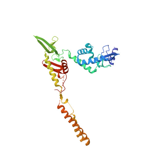
    4KIS

  • PubMed Abstract: 

    Serine integrases catalyze the integration of bacteriophage DNA into a host genome by site-specific recombination between 'attachment sites' in the phage (attP) and the host (attB). The reaction is highly directional; the reverse excision reaction between the product attL and attR sites does not occur in the absence of a phage-encoded factor, nor does recombination occur between other pairings of attachment sites. A mechanistic understanding of how these enzymes achieve site-selectivity and directionality has been limited by a lack of structural models. Here, we report the structure of the C-terminal domains of a serine integrase bound to an attP DNA half-site. The structure leads directly to models for understanding how the integrase-bound attP and attB sites differ, why these enzymes preferentially form attP × attB synaptic complexes to initiate recombination, and how attL × attR recombination is prevented. In these models, different domain organizations on attP vs. attB half-sites allow attachment-site specific interactions to form between integrase subunits via an unusual protruding coiled-coil motif. These interactions are used to preferentially synapse integrase-bound attP and attB and inhibit synapsis of integrase-bound attL and attR. The results provide a structural framework for understanding, testing and engineering serine integrase function.


  • Organizational Affiliation
    • Department of Biochemistry and Biophysics, Perelman School of Medicine, University of Pennsylvania, Philadelphia, PA 19104, USA and NE-CAT and Department of Chemistry and Chemical Biology, Cornell University, Building 436E, Argonne National Laboratory, 9700 S. Cass Avenue, Argonne, IL 60439, USA.

Macromolecule Content 

  • Total Structure Weight: 221.19 kDa 
  • Atom Count: 14,347 
  • Modeled Residue Count: 1,424 
  • Deposited Residue Count: 1,520 
  • Unique protein chains: 1
  • Unique nucleic acid chains: 2

Macromolecules


Find similar proteins by:|  3D Structure
Entity ID: 1
MoleculeChains  Sequence LengthOrganismDetailsImage
Putative integrase [Bacteriophage A118]A,
D [auth B],
G [auth C],
J [auth D]
328Listeria innocua Clip11262Mutation(s): 0 
Gene Names: int
UniProt
Find proteins for Q928V6 (Listeria innocua serovar 6a (strain ATCC BAA-680 / CLIP 11262))
Explore Q928V6 
Go to UniProtKB:  Q928V6
Entity Groups
Sequence Clusters30% Identity50% Identity70% Identity90% Identity95% Identity100% Identity
UniProt GroupQ928V6
Sequence Annotations
Expand
Reference Sequence
Find similar nucleic acids by:  (by identity cutoff) 
Entity ID: 2
MoleculeChains LengthOrganismImage
DNA (26-MER)B [auth E],
E [auth G],
H [auth I],
K
26N/A
Sequence Annotations
Expand
Reference Sequence
Find similar nucleic acids by:  (by identity cutoff) 
Entity ID: 3
MoleculeChains LengthOrganismImage
DNA (26-MER)C [auth F],
F [auth H],
I [auth J],
L
26N/A
Sequence Annotations
Expand
Reference Sequence

Small Molecules

Ligands 2 Unique
IDChains Name / Formula / InChI Key2D Diagram3D Interactions
ZN

Query on ZN


Download:Ideal Coordinates CCD File
M [auth A]
N [auth A]
O [auth A]
S [auth B]
V [auth C]
M [auth A],
N [auth A],
O [auth A],
S [auth B],
V [auth C],
X [auth D]
ZINC ION
Zn
PTFCDOFLOPIGGS-UHFFFAOYSA-N
CA

Query on CA


Download:Ideal Coordinates CCD File
P [auth A]
Q [auth A]
R [auth E]
T [auth B]
U [auth B]
P [auth A],
Q [auth A],
R [auth E],
T [auth B],
U [auth B],
W [auth C],
Y [auth D]
CALCIUM ION
Ca
BHPQYMZQTOCNFJ-UHFFFAOYSA-N

Experimental Data & Validation

Experimental Data

  • Method: X-RAY DIFFRACTION
  • Resolution: 3.20 Å
  • R-Value Free:  0.256 (Depositor), 0.250 (DCC) 
  • R-Value Work:  0.236 (Depositor), 0.219 (DCC) 
  • R-Value Observed: 0.239 (Depositor) 
Space Group: I 2 3
Unit Cell:
Length ( Å )Angle ( ˚ )
a = 290.75α = 90
b = 290.75β = 90
c = 290.75γ = 90
Software Package:
Software NamePurpose
ADSCdata collection
SOLVEphasing
CNSrefinement
HKL-2000data reduction
SCALEPACKdata scaling

Structure Validation

View Full Validation Report


Entry History 

Deposition Data

Revision History  (Full details and data files)

  • Version 1.0: 2013-07-10
    Type: Initial release
  • Version 1.1: 2013-10-23
    Changes: Database references
  • Version 1.2: 2024-02-28
    Changes: Data collection, Database references, Derived calculations