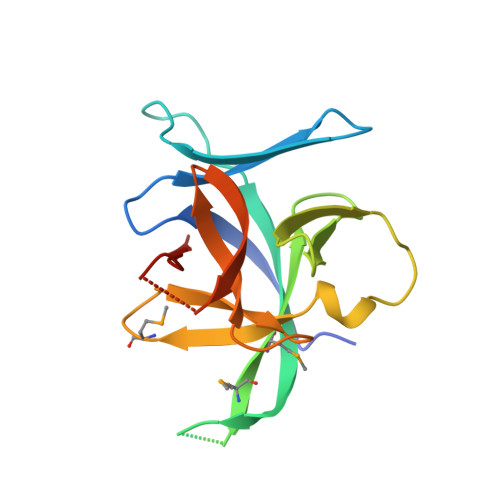
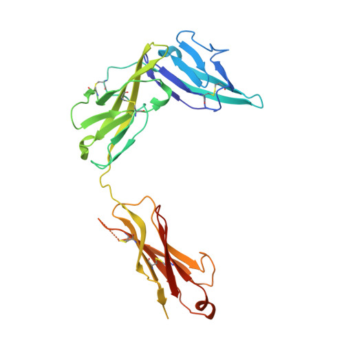
4KC3

Cytokine/receptor binary complex


Experimental Data Snapshot

  • Method: X-RAY DIFFRACTION
  • Resolution: 3.27 Å
  • R-Value Free: 0.283 
  • R-Value Work: 0.238 
  • R-Value Observed: 0.241 

Starting Models: experimental
View more details

wwPDB Validation   3D Report Full Report


Ligand Structure Quality Assessment 


This is version 1.5 of the entry. See complete history


Literature

Structural insights into the interaction of IL-33 with its receptors.

Liu, X.Hammel, M.He, Y.Tainer, J.A.Jeng, U.S.Zhang, L.Wang, S.Wang, X.

(2013) Proc Natl Acad Sci U S A 110: 14918-14923

  • DOI: https://doi.org/10.1073/pnas.1308651110
  • Primary Citation of Related Structures:  
    4KC3

  • PubMed Abstract: 

    Interleukin (IL)-33 is an important member of the IL-1 family that has pleiotropic activities in innate and adaptive immune responses in host defense and disease. It signals through its ligand-binding primary receptor ST2 and IL-1 receptor accessory protein (IL-1RAcP), both of which are members of the IL-1 receptor family. To clarify the interaction of IL-33 with its receptors, we determined the crystal structure of IL-33 in complex with the ectodomain of ST2 at a resolution of 3.27 Å. Coupled with structure-based mutagenesis and binding assay, the structural results define the molecular mechanism by which ST2 specifically recognizes IL-33. Structural comparison with other ligand-receptor complexes in the IL-1 family indicates that surface-charge complementarity is critical in determining ligand-binding specificity of IL-1 primary receptors. Combined crystallography and small-angle X-ray-scattering studies reveal that ST2 possesses hinge flexibility between the D3 domain and D1D2 module, whereas IL-1RAcP exhibits a rigid conformation in the unbound state in solution. The molecular flexibility of ST2 provides structural insights into domain-level conformational change of IL-1 primary receptors upon ligand binding, and the rigidity of IL-1RAcP explains its inability to bind ligands directly. The solution architecture of IL-33-ST2-IL-1RAcP complex from small-angle X-ray-scattering analysis resembles IL-1β-IL-1RII-IL-1RAcP and IL-1β-IL-1RI-IL-1RAcP crystal structures. The collective results confer IL-33 structure-function relationships, supporting and extending a general model for ligand-receptor assembly and activation in the IL-1 family.


  • Organizational Affiliation

    Ministry of Education Key Laboratory of Protein Science, Center for Structural Biology, School of Life Sciences, Tsinghua University, Beijing 100084, China.


Macromolecules
Find similar proteins by:  (by identity cutoff)  |  3D Structure
Entity ID: 1
MoleculeChains Sequence LengthOrganismDetailsImage
Interleukin-33159Homo sapiensMutation(s): 0 
Gene Names: IL33C9orf26IL1F11NFHEV
UniProt & NIH Common Fund Data Resources
Find proteins for O95760 (Homo sapiens)
Explore O95760 
Go to UniProtKB:  O95760
GTEx:  ENSG00000137033 
Entity Groups  
Sequence Clusters30% Identity50% Identity70% Identity90% Identity95% Identity100% Identity
UniProt GroupO95760
Sequence Annotations
Expand
  • Reference Sequence
Find similar proteins by:  (by identity cutoff)  |  3D Structure
Entity ID: 2
MoleculeChains Sequence LengthOrganismDetailsImage
Interleukin-1 receptor-like 1309Homo sapiensMutation(s): 0 
Gene Names: IL1RL1DER4ST2T1
UniProt & NIH Common Fund Data Resources
Find proteins for Q01638 (Homo sapiens)
Explore Q01638 
Go to UniProtKB:  Q01638
GTEx:  ENSG00000115602 
Entity Groups  
Sequence Clusters30% Identity50% Identity70% Identity90% Identity95% Identity100% Identity
UniProt GroupQ01638
Glycosylation
Glycosylation Sites: 3Go to GlyGen: Q01638-1
Sequence Annotations
Expand
  • Reference Sequence
Small Molecules
Modified Residues  1 Unique
IDChains TypeFormula2D DiagramParent
MSE
Query on MSE
A
L-PEPTIDE LINKINGC5 H11 N O2 SeMET
Experimental Data & Validation

Experimental Data

  • Method: X-RAY DIFFRACTION
  • Resolution: 3.27 Å
  • R-Value Free: 0.283 
  • R-Value Work: 0.238 
  • R-Value Observed: 0.241 
  • Space Group: H 3 2
Unit Cell:
Length ( Å )Angle ( ˚ )
a = 329.924α = 90
b = 329.924β = 90
c = 43.731γ = 120
Software Package:
Software NamePurpose
HKL-2000data collection
PHASERphasing
PHENIXrefinement
HKL-2000data reduction
HKL-2000data scaling

Structure Validation

View Full Validation Report


Ligand Structure Quality Assessment 


Entry History 

Deposition Data

Revision History  (Full details and data files)

  • Version 1.0: 2013-08-28
    Type: Initial release
  • Version 1.1: 2013-09-11
    Changes: Database references
  • Version 1.2: 2013-09-25
    Changes: Database references
  • Version 1.3: 2020-07-29
    Type: Remediation
    Reason: Carbohydrate remediation
    Changes: Data collection, Database references, Derived calculations, Structure summary
  • Version 1.4: 2023-09-20
    Changes: Data collection, Database references, Refinement description, Structure summary
  • Version 1.5: 2023-12-06
    Changes: Data collection