4KB9 | pdb_00004kb9

Crystal structure of wild-type HIV-1 protease with novel tricyclic P2-ligands GRL-0739A


Experimental Data Snapshot

  • Method: X-RAY DIFFRACTION
  • Resolution: 1.29 Å
  • R-Value Free: 
    0.180 (Depositor), 0.177 (DCC) 
  • R-Value Work: 
    0.139 (Depositor), 0.149 (DCC) 
  • R-Value Observed: 
    0.139 (Depositor) 

Starting Model: experimental
View more details

wwPDB Validation 3D Report Full Report

Validation slider image for 4KB9

Ligand Structure Quality Assessment 


This is version 1.2 of the entry. See complete history

Literature

Highly Potent HIV-1 Protease Inhibitors with Novel Tricyclic P2 Ligands: Design, Synthesis, and Protein-Ligand X-ray Studies.

Ghosh, A.K.Parham, G.L.Martyr, C.D.Nyalapatla, P.R.Osswald, H.L.Agniswamy, J.Wang, Y.F.Amano, M.Weber, I.T.Mitsuya, H.

(2013) J Med Chem 56: 6792-6802

  • DOI: https://doi.org/10.1021/jm400768f
  • Primary Citation Related Structures: 
    4KB9

  • PubMed Abstract: 

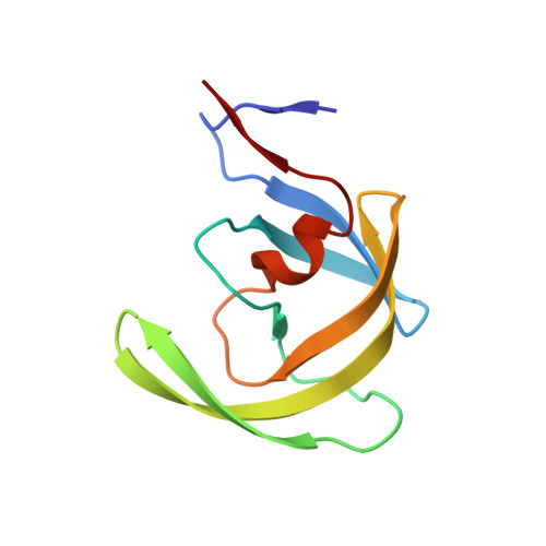
    The design, synthesis, and biological evaluation of a series of HIV-1 protease inhibitors incorporating stereochemically defined fused tricyclic P2 ligands are described. Various substituent effects were investigated to maximize the ligand-binding site interactions in the protease active site. Inhibitors 16a and 16f showed excellent enzyme inhibitory and antiviral activity, although the incorporation of sulfone functionality resulted in a decrease in potency. Both inhibitors 16a and 16f maintained activity against a panel of multidrug resistant HIV-1 variants. A high-resolution X-ray crystal structure of 16a-bound HIV-1 protease revealed important molecular insights into the ligand-binding site interactions, which may account for the inhibitor's potent antiviral activity and excellent resistance profiles.


  • Organizational Affiliation
    • Department of Chemistry and Department of Medicinal Chemistry, Purdue University , West Lafayette, Indiana 47907, United States.

Macromolecule Content 

  • Total Structure Weight: 22.55 kDa 
  • Atom Count: 1,970 
  • Modeled Residue Count: 198 
  • Deposited Residue Count: 198 
  • Unique protein chains: 1

Macromolecules

Find similar proteins by:|  3D Structure
Entity ID: 1
MoleculeChains  Sequence LengthOrganismDetailsImage
Protease
A, B
99Human immunodeficiency virus 1Mutation(s): 5 
Gene Names: pol
EC: 3.4.23.16
UniProt
Find proteins for Q7SSI0 (Human immunodeficiency virus type 1)
Explore Q7SSI0 
Go to UniProtKB:  Q7SSI0
Entity Groups
Sequence Clusters30% Identity50% Identity70% Identity90% Identity95% Identity100% Identity
UniProt GroupQ7SSI0
Sequence Annotations
Expand
Reference Sequence

Small Molecules

Ligands 5 Unique
IDChains Name / Formula / InChI Key2D Diagram3D Interactions
G79

Query on G79


Download:Ideal Coordinates CCD File
C [auth A](3aR,3bR,4S,7aR,8aS)-decahydrofuro[2,3-b][1]benzofuran-4-yl [(2S,3R)-3-hydroxy-4-{[(4-methoxyphenyl)sulfonyl](2-methylpropyl)amino}-1-phenylbutan-2-yl]carbamate
C32 H44 N2 O8 S
ORJPZJYXIFRPSW-OTTDUJIMSA-N
GOL

Query on GOL


Download:Ideal Coordinates CCD File
J [auth B],
K [auth B]
GLYCEROL
C3 H8 O3
PEDCQBHIVMGVHV-UHFFFAOYSA-N
ACT

Query on ACT


Download:Ideal Coordinates CCD File
E [auth A],
F [auth A],
I [auth B]
ACETATE ION
C2 H3 O2
QTBSBXVTEAMEQO-UHFFFAOYSA-M
CL

Query on CL


Download:Ideal Coordinates CCD File
D [auth A],
H [auth B]
CHLORIDE ION
Cl
VEXZGXHMUGYJMC-UHFFFAOYSA-M
NA

Query on NA


Download:Ideal Coordinates CCD File
G [auth B]SODIUM ION
Na
FKNQFGJONOIPTF-UHFFFAOYSA-N

Experimental Data & Validation

Experimental Data

  • Method: X-RAY DIFFRACTION
  • Resolution: 1.29 Å
  • R-Value Free:  0.180 (Depositor), 0.177 (DCC) 
  • R-Value Work:  0.139 (Depositor), 0.149 (DCC) 
  • R-Value Observed: 0.139 (Depositor) 
Space Group: P 21 21 2
Unit Cell:
Length ( Å )Angle ( ˚ )
a = 58.477α = 90
b = 86.3β = 90
c = 46.045γ = 90
Software Package:
Software NamePurpose
HKL-2000data collection
PHASESphasing
SHELXL-97refinement
HKL-2000data reduction
HKL-2000data scaling

Structure Validation

View Full Validation Report


Ligand Structure Quality Assessment 


Entry History 

Deposition Data

Revision History  (Full details and data files)

  • Version 1.0: 2013-09-04
    Type: Initial release
  • Version 1.1: 2013-09-25
    Changes: Database references
  • Version 1.2: 2023-09-20
    Changes: Data collection, Database references, Derived calculations, Refinement description