4KAG | pdb_00004kag

Crystal structure analysis of a single amino acid deletion mutation in EGFP


Experimental Data Snapshot

  • Method: X-RAY DIFFRACTION
  • Resolution: 1.12 Å
  • R-Value Free: 
    0.161 (Depositor), 0.174 (DCC) 
  • R-Value Work: 
    0.144 (Depositor), 0.153 (DCC) 
  • R-Value Observed: 
    0.145 (Depositor) 

Starting Model: experimental
View more details

wwPDB Validation 3D Report Full Report

Validation slider image for 4KAG

This is version 1.4 of the entry. See complete history

Literature

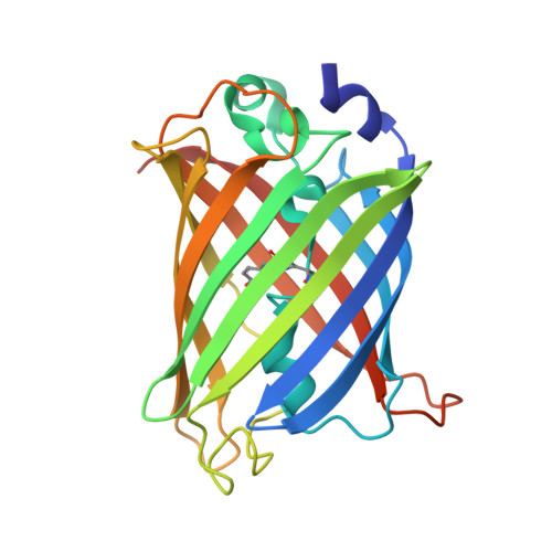
Structural and dynamic changes associated with beneficial engineered single-amino-acid deletion mutations in enhanced green fluorescent protein.

Arpino, J.A.Rizkallah, P.J.Jones, D.D.

(2014) Acta Crystallogr D Biol Crystallogr 70: 2152-2162

  • DOI: https://doi.org/10.1107/S139900471401267X
  • Primary Citation Related Structures: 
    4KAG, 4KEX

  • PubMed Abstract: 

    Single-amino-acid deletions are a common part of the natural evolutionary landscape but are rarely sampled during protein engineering owing to limited and prejudiced molecular understanding of mutations that shorten the protein backbone. Single-amino-acid deletion variants of enhanced green fluorescent protein (EGFP) have been identified by directed evolution with the beneficial effect of imparting increased cellular fluorescence. Biophysical characterization revealed that increased functional protein production and not changes to the fluorescence parameters was the mechanism that was likely to be responsible. The structure EGFP(D190Δ) containing a deletion within a loop revealed propagated changes only after the deleted residue. The structure of EGFP(A227Δ) revealed that a `flipping' mechanism was used to adjust for residue deletion at the end of a β-strand, with amino acids C-terminal to the deletion site repositioning to take the place of the deleted amino acid. In both variants new networks of short-range and long-range interactions are generated while maintaining the integrity of the hydrophobic core. Both deletion variants also displayed significant local and long-range changes in dynamics, as evident by changes in B factors compared with EGFP. Rather than being detrimental, deletion mutations can introduce beneficial structural effects through altering core protein properties, folding and dynamics, as well as function.


  • Organizational Affiliation
    • School of Biosciences, Cardiff University, Park Place, Cardiff CF10 3AT, Wales.

Macromolecule Content 

  • Total Structure Weight: 28.39 kDa 
  • Atom Count: 2,444 
  • Modeled Residue Count: 225 
  • Deposited Residue Count: 236 
  • Unique protein chains: 1

Macromolecules

Find similar proteins by:|  3D Structure
Entity ID: 1
MoleculeChains  Sequence LengthOrganismDetailsImage
Green fluorescent protein236Aequorea victoriaMutation(s): 2 
Gene Names: GFP
UniProt
Find proteins for P42212 (Aequorea victoria)
Explore P42212 
Go to UniProtKB:  P42212
Entity Groups
Sequence Clusters30% Identity50% Identity70% Identity90% Identity95% Identity100% Identity
UniProt GroupP42212
Sequence Annotations
Expand
Reference Sequence

Small Molecules

Ligands 3 Unique
IDChains Name / Formula / InChI Key2D Diagram3D Interactions
TRS

Query on TRS


Download:Ideal Coordinates CCD File
S [auth A],
T [auth A],
U [auth A]
2-AMINO-2-HYDROXYMETHYL-PROPANE-1,3-DIOL
C4 H12 N O3
LENZDBCJOHFCAS-UHFFFAOYSA-O
SO4

Query on SO4


Download:Ideal Coordinates CCD File
Q [auth A],
R [auth A]
SULFATE ION
O4 S
QAOWNCQODCNURD-UHFFFAOYSA-L
EDO

Query on EDO


Download:Ideal Coordinates CCD File
B [auth A]
C [auth A]
D [auth A]
E [auth A]
F [auth A]
B [auth A],
C [auth A],
D [auth A],
E [auth A],
F [auth A],
G [auth A],
H [auth A],
I [auth A],
J [auth A],
K [auth A],
L [auth A],
M [auth A],
N [auth A],
O [auth A],
P [auth A],
V [auth A]
1,2-ETHANEDIOL
C2 H6 O2
LYCAIKOWRPUZTN-UHFFFAOYSA-N
Modified Residues  1 Unique
IDChains TypeFormula2D DiagramParent
CRO
Query on CRO
A
L-PEPTIDE LINKINGC15 H17 N3 O5THR, TYR, GLY

Experimental Data & Validation

Experimental Data

  • Method: X-RAY DIFFRACTION
  • Resolution: 1.12 Å
  • R-Value Free:  0.161 (Depositor), 0.174 (DCC) 
  • R-Value Work:  0.144 (Depositor), 0.153 (DCC) 
  • R-Value Observed: 0.145 (Depositor) 
Space Group: P 32 2 1
Unit Cell:
Length ( Å )Angle ( ˚ )
a = 57.14α = 90
b = 57.14β = 90
c = 135.33γ = 120
Software Package:
Software NamePurpose
SCALAdata scaling
PHASERphasing
REFMACrefinement
PDB_EXTRACTdata extraction

Structure Validation

View Full Validation Report


Entry History 

Deposition Data

Revision History  (Full details and data files)

  • Version 1.0: 2014-08-06
    Type: Initial release
  • Version 1.1: 2014-09-24
    Changes: Database references
  • Version 1.2: 2023-09-20
    Changes: Data collection, Database references, Derived calculations, Refinement description
  • Version 1.3: 2023-12-06
    Changes: Data collection
  • Version 1.4: 2024-10-16
    Changes: Structure summary