4K6E | pdb_00004k6e

Crystal structure of Saccharomyces cerevisiae Dcp2 Nudix domain in complex with Mg


Experimental Data Snapshot

  • Method: X-RAY DIFFRACTION
  • Resolution: 2.10 Å
  • R-Value Free: 
    0.191 (Depositor), 0.194 (DCC) 
  • R-Value Work: 
    0.153 (Depositor), 0.154 (DCC) 
  • R-Value Observed: 
    0.157 (Depositor) 

wwPDB Validation 3D Report Full Report

Validation slider image for 4K6E

This is version 1.3 of the entry. See complete history

Literature

Active site conformational dynamics are coupled to catalysis in the mRNA decapping enzyme dcp2.

Aglietti, R.A.Floor, S.N.McClendon, C.L.Jacobson, M.P.Gross, J.D.

(2013) Structure 21: 1571-1580

  • DOI: https://doi.org/10.1016/j.str.2013.06.021
  • Primary Citation Related Structures: 
    4K6E, 4KG3, 4KG4

  • PubMed Abstract: 

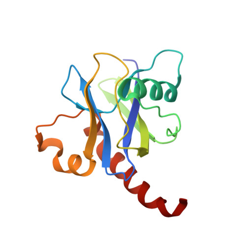
    Removal of the 5' cap structure by Dcp2 is a major step in several 5'-3' mRNA decay pathways. The activity of Dcp2 is enhanced by Dcp1 and bound coactivators, yet the details of how these interactions are linked to chemistry are poorly understood. Here, we report three crystal structures of the catalytic Nudix hydrolase domain of Dcp2 that demonstrate binding of a catalytically essential metal ion, and enzyme kinetics are used to identify several key active site residues involved in acid/base chemistry of decapping. Using nuclear magnetic resonance and molecular dynamics, we find that a conserved metal binding loop on the catalytic domain undergoes conformational changes during the catalytic cycle. These findings describe key events during the chemical step of decapping, suggest local active site conformational changes are important for activity, and provide a framework to explain stimulation of catalysis by the regulatory domain of Dcp2 and associated coactivators.


  • Organizational Affiliation
    • Program in Chemistry and Chemical Biology, University of California, San Francisco, San Francisco, CA 94158, USA; Department of Pharmaceutical Chemistry, University of California, San Francisco, San Francisco, CA 94158, USA.

Macromolecule Content 

  • Total Structure Weight: 17.14 kDa 
  • Atom Count: 1,249 
  • Modeled Residue Count: 144 
  • Deposited Residue Count: 144 
  • Unique protein chains: 1

Macromolecules

Find similar proteins by:|  3D Structure
Entity ID: 1
MoleculeChains  Sequence LengthOrganismDetailsImage
mRNA-decapping enzyme subunit 2144Saccharomyces cerevisiae S288CMutation(s): 0 
Gene Names: DCP2N1917PSU1YNL118C
EC: 3 (PDB Primary Data), 3.6.1.62 (UniProt)
UniProt
Find proteins for P53550 (Saccharomyces cerevisiae (strain ATCC 204508 / S288c))
Explore P53550 
Go to UniProtKB:  P53550
Entity Groups
Sequence Clusters30% Identity50% Identity70% Identity90% Identity95% Identity100% Identity
UniProt GroupP53550
Sequence Annotations
Expand
Reference Sequence

Small Molecules

Ligands 1 Unique
IDChains Name / Formula / InChI Key2D Diagram3D Interactions
MG

Query on MG


Download:Ideal Coordinates CCD File
B [auth A]MAGNESIUM ION
Mg
JLVVSXFLKOJNIY-UHFFFAOYSA-N

Experimental Data & Validation

Experimental Data

  • Method: X-RAY DIFFRACTION
  • Resolution: 2.10 Å
  • R-Value Free:  0.191 (Depositor), 0.194 (DCC) 
  • R-Value Work:  0.153 (Depositor), 0.154 (DCC) 
  • R-Value Observed: 0.157 (Depositor) 
Space Group: P 62
Unit Cell:
Length ( Å )Angle ( ˚ )
a = 85.054α = 90
b = 85.054β = 90
c = 48.749γ = 120
Software Package:
Software NamePurpose
DENZOdata reduction
SCALEPACKdata scaling
PHASERphasing
PHENIXrefinement
PDB_EXTRACTdata extraction

Structure Validation

View Full Validation Report


Entry History 

Deposition Data

Revision History  (Full details and data files)

  • Version 1.0: 2013-08-21
    Type: Initial release
  • Version 1.1: 2013-09-25
    Changes: Database references
  • Version 1.2: 2017-11-15
    Changes: Refinement description
  • Version 1.3: 2024-02-28
    Changes: Data collection, Database references, Derived calculations