4K50 | pdb_00004k50

Rhinovirus 16 polymerase elongation complex (r1_form)


Experimental Data Snapshot

  • Method: X-RAY DIFFRACTION
  • Resolution: 2.93 Å
  • R-Value Free: 
    0.247 (Depositor), 0.249 (DCC) 
  • R-Value Work: 
    0.191 (Depositor), 0.192 (DCC) 
  • R-Value Observed: 
    0.194 (Depositor) 

wwPDB Validation 3D Report Full Report

Validation slider image for 4K50

This is version 1.2 of the entry. See complete history

Literature

Structures of coxsackievirus, rhinovirus, and poliovirus polymerase elongation complexes solved by engineering RNA mediated crystal contacts.

Gong, P.Kortus, M.G.Nix, J.C.Davis, R.E.Peersen, O.B.

(2013) PLoS One 8: e60272-e60272

  • DOI: https://doi.org/10.1371/journal.pone.0060272
  • Primary Citation Related Structures: 
    4K4S, 4K4T, 4K4U, 4K4V, 4K4W, 4K4X, 4K4Y, 4K4Z, 4K50

  • PubMed Abstract: 

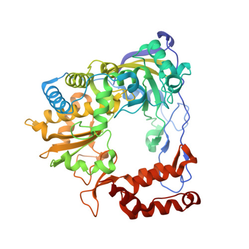
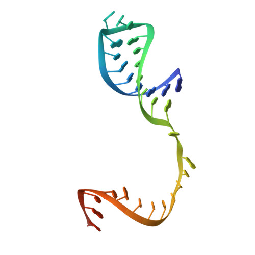
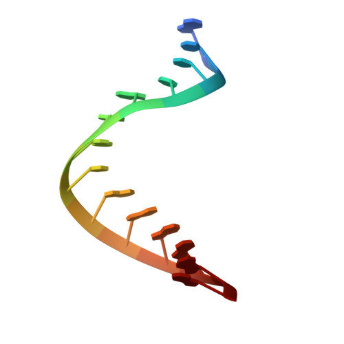
    RNA-dependent RNA polymerases play a vital role in the growth of RNA viruses where they are responsible for genome replication, but do so with rather low fidelity that allows for the rapid adaptation to different host cell environments. These polymerases are also a target for antiviral drug development. However, both drug discovery efforts and our understanding of fidelity determinants have been hampered by a lack of detailed structural information about functional polymerase-RNA complexes and the structural changes that take place during the elongation cycle. Many of the molecular details associated with nucleotide selection and catalysis were revealed in our recent structure of the poliovirus polymerase-RNA complex solved by first purifying and then crystallizing stalled elongation complexes. In the work presented here we extend that basic methodology to determine nine new structures of poliovirus, coxsackievirus, and rhinovirus elongation complexes at 2.2-2.9 Å resolution. The structures highlight conserved features of picornaviral polymerases and the interactions they make with the template and product RNA strands, including a tight grip on eight basepairs of the nascent duplex, a fully pre-positioned templating nucleotide, and a conserved binding pocket for the +2 position template strand base. At the active site we see a pre-bound magnesium ion and there is conservation of a non-standard backbone conformation of the template strand in an interaction that may aid in triggering RNA translocation via contact with the conserved polymerase motif B. Moreover, by engineering plasticity into RNA-RNA contacts, we obtain crystal forms that are capable of multiple rounds of in-crystal catalysis and RNA translocation. Together, the data demonstrate that engineering flexible RNA contacts to promote crystal lattice formation is a versatile platform that can be used to solve the structures of viral RdRP elongation complexes and their catalytic cycle intermediates.


  • Organizational Affiliation
    • Department of Biochemistry and Molecular Biology, Colorado State University, Fort Collins, Colorado, United States of America.

Macromolecule Content 

  • Total Structure Weight: 274.44 kDa 
  • Atom Count: 19,187 
  • Modeled Residue Count: 2,025 
  • Deposited Residue Count: 2,036 
  • Unique protein chains: 1
  • Unique nucleic acid chains: 2

Macromolecules


Find similar proteins by:|  3D Structure
Entity ID: 1
MoleculeChains  Sequence LengthOrganismDetailsImage
RNA polymerase 3D-POLA,
D [auth E],
G [auth I],
J [auth M]
460rhinovirus A16Mutation(s): 0 
EC: 2.7.7.48
UniProt
Find proteins for Q82122 (Human rhinovirus 16)
Explore Q82122 
Go to UniProtKB:  Q82122
Entity Groups
Sequence Clusters30% Identity50% Identity70% Identity90% Identity95% Identity100% Identity
UniProt GroupQ82122
Sequence Annotations
Expand
Reference Sequence
Find similar nucleic acids by:  (by identity cutoff) 
Entity ID: 2
MoleculeChains LengthOrganismImage
RNA (33-MER)B,
E [auth F],
H [auth J],
K [auth N]
35N/A
Sequence Annotations
Expand
Reference Sequence
Find similar nucleic acids by:  Sequence
Entity ID: 3
MoleculeChains LengthOrganismImage
RNA (5'-R(P*GP*CP*CP*CP*GP*GP*AP*CP*GP*AP*GP*AP*GP*A)-3')C,
F [auth G],
I [auth K],
L [auth O]
14N/A
Sequence Annotations
Expand
Reference Sequence

Small Molecules

Ligands 3 Unique
IDChains Name / Formula / InChI Key2D Diagram3D Interactions
SO4

Query on SO4


Download:Ideal Coordinates CCD File
P [auth A]SULFATE ION
O4 S
QAOWNCQODCNURD-UHFFFAOYSA-L
GOL

Query on GOL


Download:Ideal Coordinates CCD File
BA [auth I],
EA [auth I],
M [auth A]
GLYCEROL
C3 H8 O3
PEDCQBHIVMGVHV-UHFFFAOYSA-N
ACT

Query on ACT


Download:Ideal Coordinates CCD File
AA [auth I]
CA [auth I]
DA [auth I]
FA [auth I]
GA [auth I]
AA [auth I],
CA [auth I],
DA [auth I],
FA [auth I],
GA [auth I],
HA [auth J],
IA [auth K],
JA [auth M],
KA [auth M],
LA [auth M],
N [auth A],
O [auth A],
Q [auth A],
R [auth A],
S [auth B],
T [auth B],
U [auth E],
V [auth E],
W [auth E],
X [auth E],
Y [auth F],
Z [auth F]
ACETATE ION
C2 H3 O2
QTBSBXVTEAMEQO-UHFFFAOYSA-M

Experimental Data & Validation

Experimental Data

  • Method: X-RAY DIFFRACTION
  • Resolution: 2.93 Å
  • R-Value Free:  0.247 (Depositor), 0.249 (DCC) 
  • R-Value Work:  0.191 (Depositor), 0.192 (DCC) 
  • R-Value Observed: 0.194 (Depositor) 
Space Group: P 1
Unit Cell:
Length ( Å )Angle ( ˚ )
a = 53.75α = 90.01
b = 113.893β = 90.49
c = 122.6γ = 90
Software Package:
Software NamePurpose
d*TREKdata scaling
d*TREKdata reduction
PHENIXrefinement
PDB_EXTRACTdata extraction
Blu-Icedata collection
PHENIXphasing

Structure Validation

View Full Validation Report


Entry History 

Deposition Data

Revision History  (Full details and data files)

  • Version 1.0: 2013-05-22
    Type: Initial release
  • Version 1.1: 2013-07-03
    Changes: Database references
  • Version 1.2: 2024-02-28
    Changes: Data collection, Database references, Derived calculations