4K3C | pdb_00004k3c

The crystal structure of BamA from Haemophilus ducreyi lacking POTRA domains 1-3


Experimental Data Snapshot

  • Method: X-RAY DIFFRACTION
  • Resolution: 2.91 Å
  • R-Value Free: 
    0.270 (Depositor) 
  • R-Value Work: 
    0.220 (Depositor) 
  • R-Value Observed: 
    0.224 (Depositor) 

wwPDB Validation 3D Report Full Report

Validation slider image for 4K3C

This is version 1.3 of the entry. See complete history

Literature

Structural insight into the biogenesis of beta-barrel membrane proteins.

Noinaj, N.Kuszak, A.J.Gumbart, J.C.Lukacik, P.Chang, H.Easley, N.C.Lithgow, T.Buchanan, S.K.

(2013) Nature 501: 385-390

  • DOI: https://doi.org/10.1038/nature12521
  • Primary Citation Related Structures: 
    4K3B, 4K3C

  • PubMed Abstract: 

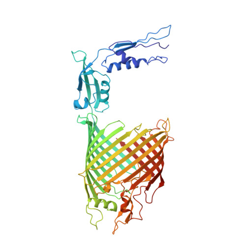
    β-barrel membrane proteins are essential for nutrient import, signalling, motility and survival. In Gram-negative bacteria, the β-barrel assembly machinery (BAM) complex is responsible for the biogenesis of β-barrel membrane proteins, with homologous complexes found in mitochondria and chloroplasts. Here we describe the structure of BamA, the central and essential component of the BAM complex, from two species of bacteria: Neisseria gonorrhoeae and Haemophilus ducreyi. BamA consists of a large periplasmic domain attached to a 16-strand transmembrane β-barrel domain. Three structural features shed light on the mechanism by which BamA catalyses β-barrel assembly. First, the interior cavity is accessible in one BamA structure and conformationally closed in the other. Second, an exterior rim of the β-barrel has a distinctly narrowed hydrophobic surface, locally destabilizing the outer membrane. And third, the β-barrel can undergo lateral opening, suggesting a route from the interior cavity in BamA into the outer membrane.


  • Organizational Affiliation
    • National Institute of Diabetes and Digestive and Kidney Diseases, National Institutes of Health, Bethesda, Maryland 20892, USA.

Macromolecule Content 

  • Total Structure Weight: 59.43 kDa 
  • Atom Count: 3,959 
  • Modeled Residue Count: 532 
  • Deposited Residue Count: 532 
  • Unique protein chains: 1

Macromolecules

Find similar proteins by:|  3D Structure
Entity ID: 1
MoleculeChains  Sequence LengthOrganismDetailsImage
Outer membrane protein assembly factor BamA532[Haemophilus] ducreyiMutation(s): 0 
Gene Names: D15bamA
Membrane Entity: Yes 
UniProt
Find proteins for Q93PM2 (Haemophilus ducreyi)
Explore Q93PM2 
Go to UniProtKB:  Q93PM2
Entity Groups
Sequence Clusters30% Identity50% Identity70% Identity90% Identity95% Identity100% Identity
UniProt GroupQ93PM2
Sequence Annotations
Expand
Reference Sequence

Experimental Data & Validation

Experimental Data

  • Method: X-RAY DIFFRACTION
  • Resolution: 2.91 Å
  • R-Value Free:  0.270 (Depositor) 
  • R-Value Work:  0.220 (Depositor) 
  • R-Value Observed: 0.224 (Depositor) 
Space Group: P 2 21 21
Unit Cell:
Length ( Å )Angle ( ˚ )
a = 47.779α = 90
b = 88.162β = 90
c = 244.454γ = 90
Software Package:
Software NamePurpose
SERGUIdata collection
PHENIX/AutoSOLmodel building
PHENIXrefinement
HKL-2000data reduction
HKL-2000data scaling
PHENIX/AutoSOLphasing

Structure Validation

View Full Validation Report


Entry History 

Deposition Data

Revision History  (Full details and data files)

  • Version 1.0: 2013-09-04
    Type: Initial release
  • Version 1.1: 2013-09-11
    Changes: Database references
  • Version 1.2: 2013-10-02
    Changes: Database references
  • Version 1.3: 2024-02-28
    Changes: Data collection, Database references