4JZK | pdb_00004jzk

Crystal Structure of Adenylate kinase of E. Coli with ADP/AMP bound


Experimental Data Snapshot

  • Method: X-RAY DIFFRACTION
  • Resolution: 1.63 Å
  • R-Value Free: 
    0.252 (Depositor), 0.253 (DCC) 
  • R-Value Work: 
    0.214 (Depositor), 0.218 (DCC) 
  • R-Value Observed: 
    0.216 (Depositor) 

wwPDB Validation 3D Report Full Report

Validation slider image for 4JZK

Ligand Structure Quality Assessment 


This is version 1.1 of the entry. See complete history

Literature

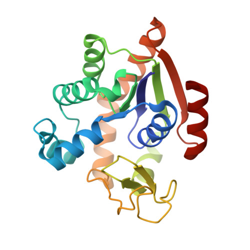
Crystal structure of E. Coli Adenylate kinase with ADP and AMP bound

Cho, Y.-J.Agafonov, R.Kern, D.

To be published.

Macromolecule Content 

  • Total Structure Weight: 48.79 kDa 
  • Atom Count: 4,109 
  • Modeled Residue Count: 428 
  • Deposited Residue Count: 428 
  • Unique protein chains: 1

Macromolecules

Find similar proteins by:|  3D Structure
Entity ID: 1
MoleculeChains  Sequence LengthOrganismDetailsImage
Adenylate kinase
A, B
214Escherichia coli O104:H4 str. 2009EL-2071Mutation(s): 0 
Gene Names: adkO3O_06175
EC: 2.7.4.3

Experimental Data & Validation

Experimental Data

  • Method: X-RAY DIFFRACTION
  • Resolution: 1.63 Å
  • R-Value Free:  0.252 (Depositor), 0.253 (DCC) 
  • R-Value Work:  0.214 (Depositor), 0.218 (DCC) 
  • R-Value Observed: 0.216 (Depositor) 
Space Group: P 21 2 21
Unit Cell:
Length ( Å )Angle ( ˚ )
a = 72.96α = 90
b = 78.8β = 90
c = 83.03γ = 90
Software Package:
Software NamePurpose
PHENIXrefinement
PDB_EXTRACTdata extraction

Structure Validation

View Full Validation Report


Ligand Structure Quality Assessment 


Entry History 

Deposition Data

Revision History  (Full details and data files)

  • Version 1.0: 2014-04-30
    Type: Initial release
  • Version 1.1: 2024-02-28
    Changes: Data collection, Database references, Derived calculations