4JVY | pdb_00004jvy

Structure of the STAR (signal transduction and activation of RNA) domain of GLD-1 bound to RNA


Experimental Data Snapshot

  • Method: X-RAY DIFFRACTION
  • Resolution: 2.85 Å
  • R-Value Free: 
    0.222 (Depositor), 0.220 (DCC) 
  • R-Value Work: 
    0.175 (Depositor), 0.177 (DCC) 
  • R-Value Observed: 
    0.180 (Depositor) 

wwPDB Validation 3D Report Full Report

Validation slider image for 4JVY

This is version 1.2 of the entry. See complete history

Literature

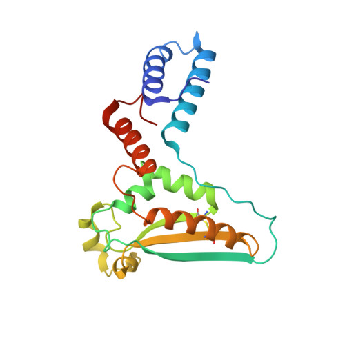
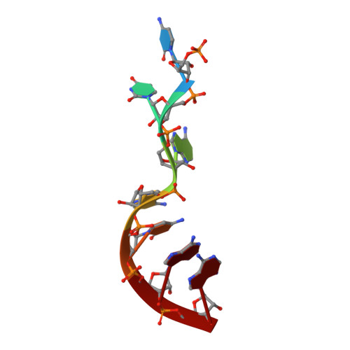
Structure-function studies of STAR family Quaking proteins bound to their in vivo RNA target sites.

Teplova, M.Hafner, M.Teplov, D.Essig, K.Tuschl, T.Patel, D.J.

(2013) Genes Dev 27: 928-940

  • DOI: https://doi.org/10.1101/gad.216531.113
  • Primary Citation Related Structures: 
    4JVH, 4JVY

  • PubMed Abstract: 

    Mammalian Quaking (QKI) and its Caenorhabditis elegans homolog, GLD-1 (defective in germ line development), are evolutionarily conserved RNA-binding proteins, which post-transcriptionally regulate target genes essential for developmental processes and myelination. We present X-ray structures of the STAR (signal transduction and activation of RNA) domain, composed of Qua1, K homology (KH), and Qua2 motifs of QKI and GLD-1 bound to high-affinity in vivo RNA targets containing YUAAY RNA recognition elements (RREs). The KH and Qua2 motifs of the STAR domain synergize to specifically interact with bases and sugar-phosphate backbones of the bound RRE. Qua1-mediated homodimerization generates a scaffold that enables concurrent recognition of two RREs, thereby plausibly targeting tandem RREs present in many QKI-targeted transcripts. Structure-guided mutations reduced QKI RNA-binding affinity in vitro and in vivo, and expression of QKI mutants in human embryonic kidney cells (HEK293) significantly decreased the abundance of QKI target mRNAs. Overall, our studies define principles underlying RNA target selection by STAR homodimers and provide insights into the post-transcriptional regulatory function of mammalian QKI proteins.


  • Organizational Affiliation
    • Structural Biology Program, Memorial Sloan-Kettering Cancer Center, New York, New York 10065, USA.

Macromolecule Content 

  • Total Structure Weight: 49.34 kDa 
  • Atom Count: 3,393 
  • Modeled Residue Count: 395 
  • Deposited Residue Count: 406 
  • Unique protein chains: 1
  • Unique nucleic acid chains: 1

Macromolecules


Find similar proteins by:|  3D Structure
Entity ID: 1
MoleculeChains  Sequence LengthOrganismDetailsImage
Female germline-specific tumor suppressor gld-1
A, B
196Caenorhabditis elegansMutation(s): 0 
Gene Names: gld-1T23G11.3
UniProt
Find proteins for Q17339 (Caenorhabditis elegans)
Explore Q17339 
Go to UniProtKB:  Q17339
Entity Groups
Sequence Clusters30% Identity50% Identity70% Identity90% Identity95% Identity100% Identity
UniProt GroupQ17339
Sequence Annotations
Expand
Reference Sequence
Find similar nucleic acids by:  Sequence
Entity ID: 2
MoleculeChains LengthOrganismImage
RNA (5'-R(P*CP*UP*AP*AP*CP*AP*A)-3')C [auth D],
D [auth F]
7N/A
Sequence Annotations
Expand
Reference Sequence

Experimental Data & Validation

Experimental Data

  • Method: X-RAY DIFFRACTION
  • Resolution: 2.85 Å
  • R-Value Free:  0.222 (Depositor), 0.220 (DCC) 
  • R-Value Work:  0.175 (Depositor), 0.177 (DCC) 
  • R-Value Observed: 0.180 (Depositor) 
Space Group: P 31 2 1
Unit Cell:
Length ( Å )Angle ( ˚ )
a = 142.274α = 90
b = 142.274β = 90
c = 76.652γ = 120
Software Package:
Software NamePurpose
DENZOdata reduction
SCALEPACKdata scaling
PHENIXrefinement
PDB_EXTRACTdata extraction
CBASSdata collection
AMoREphasing

Structure Validation

View Full Validation Report


Entry History 

Deposition Data

Revision History  (Full details and data files)

  • Version 1.0: 2013-05-08
    Type: Initial release
  • Version 1.1: 2017-11-15
    Changes: Advisory, Refinement description
  • Version 1.2: 2024-11-06
    Changes: Advisory, Data collection, Database references, Structure summary