4JR3

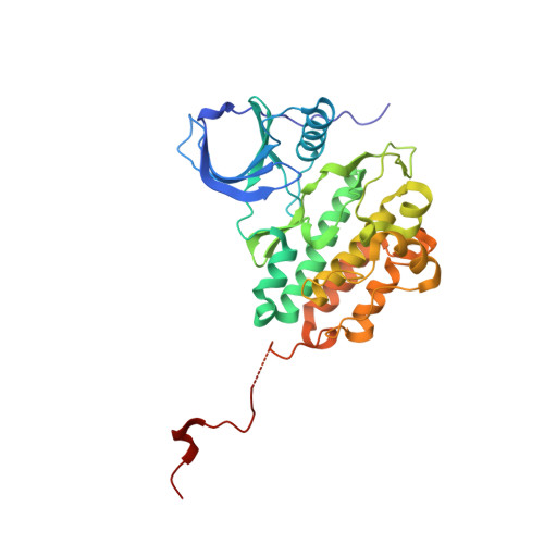
Crystal structure of EGFR kinase domain in complex with compound 3g


Experimental Data Snapshot

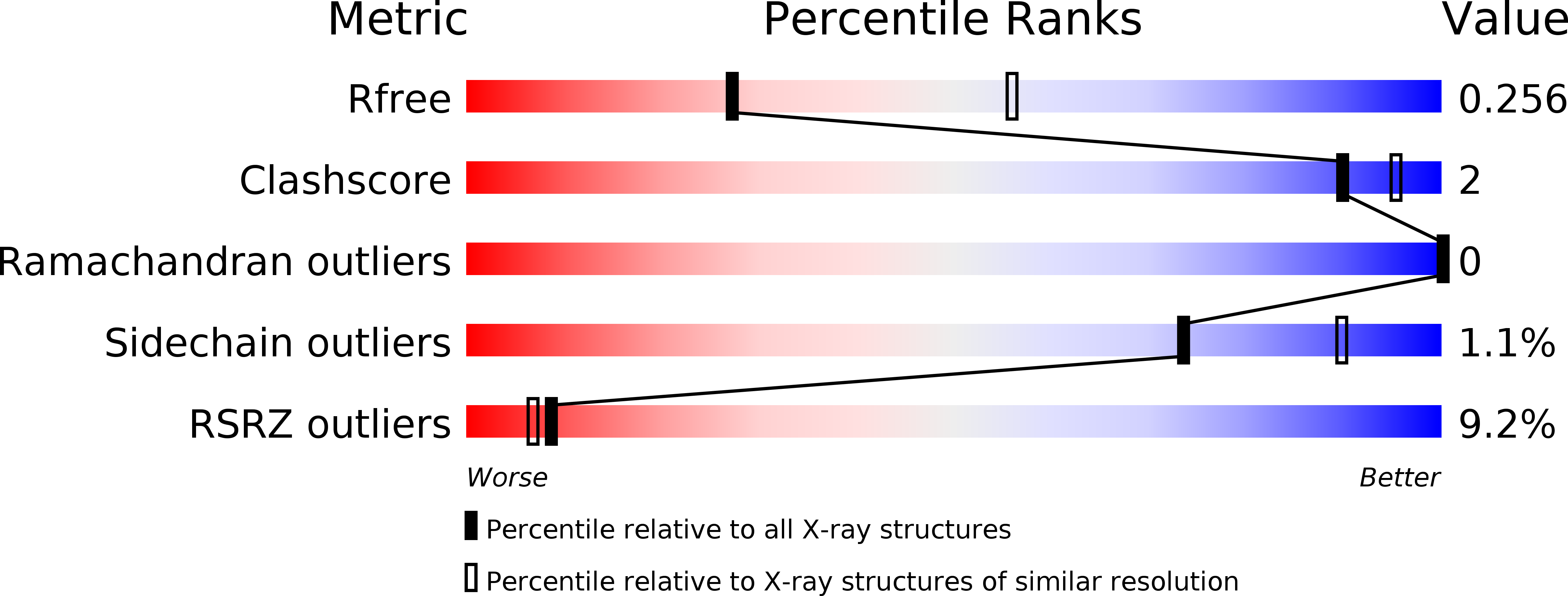
  • Method: X-RAY DIFFRACTION
  • Resolution: 2.70 Å
  • R-Value Free: 0.248 
  • R-Value Work: 0.213 
  • R-Value Observed: 0.215 

Starting Model: experimental
View more details

wwPDB Validation   3D Report Full Report


Ligand Structure Quality Assessment 


This is version 1.1 of the entry. See complete history


Literature

Protein Kinase Inhibitor Design by Targeting the Asp-Phe-Gly (DFG) Motif: The Role of the DFG Motif in the Design of Epidermal Growth Factor Receptor Inhibitors

Peng, Y.H.Shiao, H.Y.Tu, C.H.Liu, P.M.Hsu, J.T.Amancha, P.K.Wu, J.S.Coumar, M.S.Chen, C.H.Wang, S.Y.Lin, W.H.Sun, H.Y.Chao, Y.S.Lyu, P.C.Hsieh, H.P.Wu, S.Y.

(2013) J Med Chem 56: 3889-3903

  • DOI: https://doi.org/10.1021/jm400072p
  • Primary Citation of Related Structures:  
    4JQ7, 4JQ8, 4JR3, 4JRV

  • PubMed Abstract: 

    The Asp-Phe-Gly (DFG) motif plays an important role in the regulation of kinase activity. Structure-based drug design was performed to design compounds able to interact with the DFG motif; epidermal growth factor receptor (EGFR) was selected as an example. Structural insights obtained from the EGFR/2a complex suggested that an extension from the meta-position on the phenyl group (ring-5) would improve interactions with the DFG motif. Indeed, introduction of an N,N-dimethylamino tail resulted in 4b, which showed almost 50-fold improvement in inhibition compared to 2a. Structural studies confirmed this N,N-dimethylamino tail moved toward the DFG motif to form a salt bridge with the side chain of Asp831. That the interactions with the DFG motif greatly contribute to the potency of 4b is strongly evidenced by synthesizing and testing compounds 2a, 3g, and 4f: when the charge interactions are absent, the inhibitory activity decreased significantly.


  • Organizational Affiliation

    Institute of Biotechnology and Pharmaceutical Research, National Health Research Institutes, 35 Keyan Road, Zhunan Town, Miaoli County 350, Taiwan, ROC.


Macromolecules
Find similar proteins by:  (by identity cutoff)  |  3D Structure
Entity ID: 1
MoleculeChains Sequence LengthOrganismDetailsImage
Epidermal growth factor receptor328Homo sapiensMutation(s): 0 
Gene Names: EGFR
EC: 2.7.10.1
UniProt & NIH Common Fund Data Resources
Find proteins for P00533 (Homo sapiens)
Explore P00533 
Go to UniProtKB:  P00533
PHAROS:  P00533
GTEx:  ENSG00000146648 
Entity Groups  
Sequence Clusters30% Identity50% Identity70% Identity90% Identity95% Identity100% Identity
UniProt GroupP00533
Sequence Annotations
Expand
  • Reference Sequence
Small Molecules
Ligands 1 Unique
IDChains Name / Formula / InChI Key2D Diagram3D Interactions
KJR
Query on KJR

Download Ideal Coordinates CCD File 
B [auth A]N-[3-(4-{[(1S)-2-hydroxy-1-phenylethyl]amino}-6-phenylfuro[2,3-d]pyrimidin-5-yl)phenyl]acetamide
C28 H24 N4 O3
QTVUEQZXBRDKNW-HSZRJFAPSA-N
Binding Affinity Annotations 
IDSourceBinding Affinity
KJR PDBBind:  4JR3 IC50: 218 (nM) from 1 assay(s)
Experimental Data & Validation

Experimental Data

  • Method: X-RAY DIFFRACTION
  • Resolution: 2.70 Å
  • R-Value Free: 0.248 
  • R-Value Work: 0.213 
  • R-Value Observed: 0.215 
  • Space Group: I 2 3
Unit Cell:
Length ( Å )Angle ( ˚ )
a = 145.69α = 90
b = 145.69β = 90
c = 145.69γ = 90
Software Package:
Software NamePurpose
HKL-2000data collection
MOLREPphasing
REFMACrefinement
HKL-2000data reduction
SCALEPACKdata scaling

Structure Validation

View Full Validation Report


Ligand Structure Quality Assessment 


Entry History 

Deposition Data

Revision History  (Full details and data files)

  • Version 1.0: 2013-06-19
    Type: Initial release
  • Version 1.1: 2023-11-08
    Changes: Data collection, Database references, Derived calculations, Refinement description