4JL6 | pdb_00004jl6

Crystal Structure of Adenylate kinase with 2 ADP's


Experimental Data Snapshot

  • Method: X-RAY DIFFRACTION
  • Resolution: 1.65 Å
  • R-Value Free: 
    0.241 (Depositor), 0.237 (DCC) 
  • R-Value Work: 
    0.208 (Depositor), 0.205 (DCC) 
  • R-Value Observed: 
    0.210 (Depositor) 

wwPDB Validation 3D Report Full Report

Validation slider image for 4JL6

Ligand Structure Quality Assessment 


This is version 1.3 of the entry. See complete history

Literature

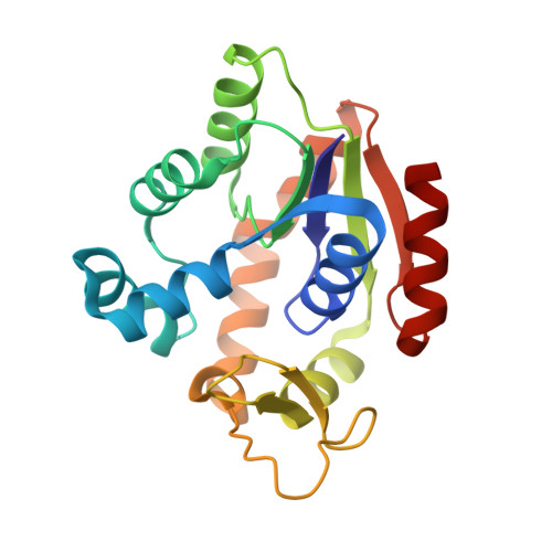
The energy landscape of adenylate kinase during catalysis.

Kerns, S.J.Agafonov, R.V.Cho, Y.J.Pontiggia, F.Otten, R.Pachov, D.V.Kutter, S.Phung, L.A.Murphy, P.N.Thai, V.Alber, T.Hagan, M.F.Kern, D.

(2015) Nat Struct Mol Biol 22: 124-131

  • DOI: https://doi.org/10.1038/nsmb.2941
  • Primary Citation Related Structures: 
    3SR0, 4CF7, 4JKY, 4JL5, 4JL6, 4JL8, 4JLA, 4JLB, 4JLD, 4JLO, 4JLP

  • PubMed Abstract: 

    Kinases perform phosphoryl-transfer reactions in milliseconds; without enzymes, these reactions would take about 8,000 years under physiological conditions. Despite extensive studies, a comprehensive understanding of kinase energy landscapes, including both chemical and conformational steps, is lacking. Here we scrutinize the microscopic steps in the catalytic cycle of adenylate kinase, through a combination of NMR measurements during catalysis, pre-steady-state kinetics, molecular-dynamics simulations and crystallography of active complexes. We find that the Mg(2+) cofactor activates two distinct molecular events: phosphoryl transfer (>10(5)-fold) and lid opening (10(3)-fold). In contrast, mutation of an essential active site arginine decelerates phosphoryl transfer 10(3)-fold without substantially affecting lid opening. Our results highlight the importance of the entire energy landscape in catalysis and suggest that adenylate kinases have evolved to activate key processes simultaneously by precise placement of a single, charged and very abundant cofactor in a preorganized active site.


  • Organizational Affiliation
    • Howard Hughes Medical Institute, Department of Biochemistry, Brandeis University, Waltham, Massachusetts, USA.

Macromolecule Content 

  • Total Structure Weight: 47.67 kDa 
  • Atom Count: 3,834 
  • Modeled Residue Count: 406 
  • Deposited Residue Count: 406 
  • Unique protein chains: 1

Macromolecules

Find similar proteins by:|  3D Structure
Entity ID: 1
MoleculeChains  Sequence LengthOrganismDetailsImage
Adenylate kinase
A, B
203Aquifex aeolicus VF5Mutation(s): 0 
Gene Names: adkaq_078
EC: 2.7.4.3
UniProt
Find proteins for O66490 (Aquifex aeolicus (strain VF5))
Explore O66490 
Go to UniProtKB:  O66490
Entity Groups
Sequence Clusters30% Identity50% Identity70% Identity90% Identity95% Identity100% Identity
UniProt GroupO66490
Sequence Annotations
Expand
Reference Sequence

Experimental Data & Validation

Experimental Data

  • Method: X-RAY DIFFRACTION
  • Resolution: 1.65 Å
  • R-Value Free:  0.241 (Depositor), 0.237 (DCC) 
  • R-Value Work:  0.208 (Depositor), 0.205 (DCC) 
  • R-Value Observed: 0.210 (Depositor) 
Space Group: P 21 21 21
Unit Cell:
Length ( Å )Angle ( ˚ )
a = 66.47α = 90
b = 71.56β = 90
c = 86.3γ = 90
Software Package:
Software NamePurpose
PHENIXrefinement
PDB_EXTRACTdata extraction
MOSFLMdata reduction
SCALAdata scaling
PHASERphasing

Structure Validation

View Full Validation Report


Ligand Structure Quality Assessment 


Entry History 

Deposition Data

Revision History  (Full details and data files)

  • Version 1.0: 2014-06-25
    Type: Initial release
  • Version 1.1: 2015-01-28
    Changes: Database references
  • Version 1.2: 2015-02-18
    Changes: Database references
  • Version 1.3: 2024-02-28
    Changes: Data collection, Database references, Derived calculations