4JKV | pdb_00004jkv

Structure of the human smoothened 7TM receptor in complex with an antitumor agent


Experimental Data Snapshot

  • Method: X-RAY DIFFRACTION
  • Resolution: 2.45 Å
  • R-Value Free: 
    0.231 (Depositor) 
  • R-Value Work: 
    0.200 (Depositor) 
  • R-Value Observed: 
    0.201 (Depositor) 

Starting Model: experimental
View more details

wwPDB Validation 3D Report Full Report

Validation slider image for 4JKV

Ligand Structure Quality Assessment 


This is version 1.7 of the entry. See complete history

Literature

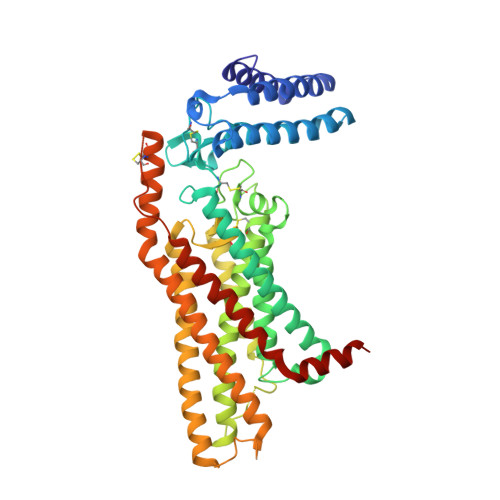
Structure of the human smoothened receptor bound to an antitumour agent.

Wang, C.Wu, H.Katritch, V.Han, G.W.Huang, X.P.Liu, W.Siu, F.Y.Roth, B.L.Cherezov, V.Stevens, R.C.

(2013) Nature 497: 338-343

  • DOI: https://doi.org/10.1038/nature12167
  • Primary Citation Related Structures: 
    4JKV

  • PubMed Abstract: 

    The smoothened (SMO) receptor, a key signal transducer in the hedgehog signalling pathway, is responsible for the maintenance of normal embryonic development and is implicated in carcinogenesis. It is classified as a class frizzled (class F) G-protein-coupled receptor (GPCR), although the canonical hedgehog signalling pathway involves the GLI transcription factors and the sequence similarity with class A GPCRs is less than 10%. Here we report the crystal structure of the transmembrane domain of the human SMO receptor bound to the small-molecule antagonist LY2940680 at 2.5 Å resolution. Although the SMO receptor shares the seven-transmembrane helical fold, most of the conserved motifs for class A GPCRs are absent, and the structure reveals an unusually complex arrangement of long extracellular loops stabilized by four disulphide bonds. The ligand binds at the extracellular end of the seven-transmembrane-helix bundle and forms extensive contacts with the loops.


  • Organizational Affiliation
    • Department of Integrative Structural and Computational Biology, The Scripps Research Institute, 10550 North Torrey Pines Road, La Jolla, California 92037, USA.

Macromolecule Content 

  • Total Structure Weight: 110.45 kDa 
  • Atom Count: 7,390 
  • Modeled Residue Count: 904 
  • Deposited Residue Count: 950 
  • Unique protein chains: 1

Macromolecules

Find similar proteins by:|  3D Structure
Entity ID: 1
MoleculeChains  Sequence LengthOrganismDetailsImage
Soluble cytochrome b562, Smoothened homolog
A, B
475Escherichia coliHomo sapiens
This entity is chimeric
Mutation(s): 2 
Gene Names: cybCSMO_HUMANSMOSMOH
Membrane Entity: Yes 
UniProt & NIH Common Fund Data Resources
Find proteins for P0ABE7 (Escherichia coli)
Explore P0ABE7 
Go to UniProtKB:  P0ABE7
Find proteins for Q99835 (Homo sapiens)
Explore Q99835 
Go to UniProtKB:  Q99835
PHAROS:  Q99835
GTEx:  ENSG00000128602 
Entity Groups
Sequence Clusters30% Identity50% Identity70% Identity90% Identity95% Identity100% Identity
UniProt GroupsP0ABE7Q99835
Sequence Annotations
Expand
Reference Sequence

Small Molecules

Ligands 6 Unique
IDChains Name / Formula / InChI Key2D Diagram3D Interactions
1KS

Query on 1KS


Download:Ideal Coordinates CCD File
C [auth A],
L [auth B]
4-fluoro-N-methyl-N-{1-[4-(1-methyl-1H-pyrazol-5-yl)phthalazin-1-yl]piperidin-4-yl}-2-(trifluoromethyl)benzamide
C26 H24 F4 N6 O
SZBGQDXLNMELTB-UHFFFAOYSA-N
OLC

Query on OLC


Download:Ideal Coordinates CCD File
D [auth A],
E [auth A],
F [auth A],
M [auth B],
N [auth B]
(2R)-2,3-dihydroxypropyl (9Z)-octadec-9-enoate
C21 H40 O4
RZRNAYUHWVFMIP-GDCKJWNLSA-N
OLA

Query on OLA


Download:Ideal Coordinates CCD File
G [auth A],
H [auth A],
O [auth B]
OLEIC ACID
C18 H34 O2
ZQPPMHVWECSIRJ-KTKRTIGZSA-N
PG4

Query on PG4


Download:Ideal Coordinates CCD File
K [auth A]TETRAETHYLENE GLYCOL
C8 H18 O5
UWHCKJMYHZGTIT-UHFFFAOYSA-N
PGE

Query on PGE


Download:Ideal Coordinates CCD File
J [auth A]TRIETHYLENE GLYCOL
C6 H14 O4
ZIBGPFATKBEMQZ-UHFFFAOYSA-N
PEG

Query on PEG


Download:Ideal Coordinates CCD File
I [auth A],
P [auth B]
DI(HYDROXYETHYL)ETHER
C4 H10 O3
MTHSVFCYNBDYFN-UHFFFAOYSA-N

Experimental Data & Validation

Experimental Data

  • Method: X-RAY DIFFRACTION
  • Resolution: 2.45 Å
  • R-Value Free:  0.231 (Depositor) 
  • R-Value Work:  0.200 (Depositor) 
  • R-Value Observed: 0.201 (Depositor) 
Space Group: P 1 21 1
Unit Cell:
Length ( Å )Angle ( ˚ )
a = 75.444α = 90
b = 98.177β = 103.27
c = 84.293γ = 90
Software Package:
Software NamePurpose
Blu-Icedata collection
AutoSolphasing
BUSTERrefinement
HKL-2000data reduction
HKL-2000data scaling

Structure Validation

View Full Validation Report


Ligand Structure Quality Assessment 


Entry History 

Deposition Data

Revision History  (Full details and data files)

  • Version 1.0: 2013-04-24
    Type: Initial release
  • Version 1.1: 2013-05-15
    Changes: Database references
  • Version 1.2: 2013-05-29
    Changes: Database references
  • Version 1.3: 2013-11-27
    Changes: Structure summary
  • Version 1.4: 2017-08-02
    Changes: Refinement description, Source and taxonomy
  • Version 1.5: 2017-11-15
    Changes: Refinement description
  • Version 1.6: 2024-04-03
    Changes: Data collection, Database references, Derived calculations, Refinement description
  • Version 1.7: 2024-11-06
    Changes: Structure summary