4JKI | pdb_00004jki

Crystal Structure of N10-Formyltetrahydrofolate Synthetase with ZD9331, Formylphosphate, and ADP


Experimental Data Snapshot

  • Method: X-RAY DIFFRACTION
  • Resolution: 2.60 Å
  • R-Value Free: 
    0.249 (Depositor), 0.231 (DCC) 
  • R-Value Work: 
    0.186 (Depositor) 
  • R-Value Observed: 
    0.190 (Depositor) 

Starting Model: experimental
View more details

wwPDB Validation 3D Report Full Report

Validation slider image for 4JKI

Ligand Structure Quality Assessment 


This is version 1.3 of the entry. See complete history

Literature

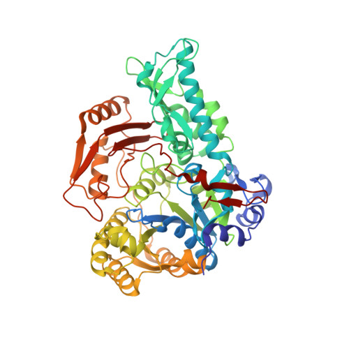
Mechanism of N10-formyltetrahydrofolate synthetase derived from complexes with intermediates and inhibitors.

Celeste, L.R.Chai, G.Bielak, M.Minor, W.Lovelace, L.L.Lebioda, L.

(2012) Protein Sci 21: 219-228

  • DOI: https://doi.org/10.1002/pro.2005
  • Primary Citation Related Structures: 
    4JIM, 4JJK, 4JJZ, 4JKI

  • PubMed Abstract: 

    N(10) -formyltetrahydrofolate synthetase (FTHFS) is a folate enzyme that catalyzes the formylation of tetrahydrofolate (THF) in an ATP dependent manner. Structures of FTHFS from the thermophilic homoacetogen, Moorella thermoacetica, complexed with (1) a catalytic intermediate-formylphosphate (XPO) and product-ADP; (2) with an inhibitory substrate analog-folate; (3) with XPO and an inhibitory THF analog, ZD9331, were used to analyze the enzyme mechanism. Nucleophilic attack of the formate ion on the gamma phosphate of ATP leads to the formation of XPO and the first product ADP. A channel that leads to the putative formate binding pocket allows for the binding of ATP and formate in random order. Formate binding is due to interactions with the gamma-phosphate moiety of ATP and additionally to two hydrogen bonds from the backbone nitrogen of Ala276 and the side chain of Arg97. Upon ADP dissociation, XPO reorients and moves to the position previously occupied by the beta-phosphate of ATP. Conformational changes that occur due to the XPO presence apparently allow for the recruitment of the third substrate, THF, with its pterin moiety positioned between Phe384 and Trp412. This position overlaps with that of the bound nucleoside, which is consistent with a catalytic mechanism hypothesis that FTHFS works via a sequential ping-pong mechanism. More specifically, a random bi uni uni bi ping-pong ter ter mechanism is proposed. Additionally, the native structure originally reported at a 2.5 Å resolution was redetermined at a 2.2 Å resolution.


  • Organizational Affiliation
    • Department of Chemistry and Biochemistry, University of South Carolina, 631 Sumter St., Columbia, SC 29208, USA.

Macromolecule Content 

  • Total Structure Weight: 121.2 kDa 
  • Atom Count: 8,377 
  • Modeled Residue Count: 1,112 
  • Deposited Residue Count: 1,118 
  • Unique protein chains: 1

Macromolecules

Find similar proteins by:|  3D Structure
Entity ID: 1
MoleculeChains  Sequence LengthOrganismDetailsImage
Formate--tetrahydrofolate ligase
A, B
559Moorella thermoacetica ATCC 39073Mutation(s): 0 
Gene Names: fhsmoorellaMoth_0109
EC: 6.3.4.3
UniProt
Find proteins for Q2RM91 (Moorella thermoacetica (strain ATCC 39073 / JCM 9320))
Explore Q2RM91 
Go to UniProtKB:  Q2RM91
Entity Groups
Sequence Clusters30% Identity50% Identity70% Identity90% Identity95% Identity100% Identity
UniProt GroupQ2RM91
Sequence Annotations
Expand
Reference Sequence

Small Molecules

Ligands 4 Unique
IDChains Name / Formula / InChI Key2D Diagram3D Interactions
ADP

Query on ADP


Download:Ideal Coordinates CCD File
E [auth A]ADENOSINE-5'-DIPHOSPHATE
C10 H15 N5 O10 P2
XTWYTFMLZFPYCI-KQYNXXCUSA-N
ZD9

Query on ZD9


Download:Ideal Coordinates CCD File
C [auth A]2,7-dimethyl-6-[(prop-1-yn-1-ylamino)methyl]quinazolin-4(3H)-one
C14 H15 N3 O
VHJQNTIPWBTKDM-UHFFFAOYSA-N
XPO

Query on XPO


Download:Ideal Coordinates CCD File
D [auth A]formyl phosphate
C H3 O5 P
TVISEJUYYBUVNV-UHFFFAOYSA-N
SO4

Query on SO4


Download:Ideal Coordinates CCD File
F [auth A],
G [auth A],
H [auth B],
I [auth B]
SULFATE ION
O4 S
QAOWNCQODCNURD-UHFFFAOYSA-L

Experimental Data & Validation

Experimental Data

  • Method: X-RAY DIFFRACTION
  • Resolution: 2.60 Å
  • R-Value Free:  0.249 (Depositor), 0.231 (DCC) 
  • R-Value Work:  0.186 (Depositor) 
  • R-Value Observed: 0.190 (Depositor) 
Space Group: H 3 2
Unit Cell:
Length ( Å )Angle ( ˚ )
a = 162.373α = 90
b = 162.373β = 90
c = 258.074γ = 120
Software Package:
Software NamePurpose
DENZOdata reduction
SCALEPACKdata scaling
REFMACrefinement
PDB_EXTRACTdata extraction
HKL-2000data collection
HKL-2000data reduction
HKL-2000data scaling
MOLREPphasing

Structure Validation

View Full Validation Report


Ligand Structure Quality Assessment 


Entry History 

Deposition Data

Revision History  (Full details and data files)

  • Version 1.0: 2013-03-20
    Type: Initial release
  • Version 1.1: 2017-11-15
    Changes: Refinement description
  • Version 1.2: 2020-09-09
    Changes: Derived calculations, Structure summary
  • Version 1.3: 2023-09-20
    Changes: Data collection, Database references, Refinement description