4JE5 | pdb_00004je5

Crystal structure of the aromatic aminotransferase Aro8, a putative alpha-aminoadipate aminotransferase in Saccharomyces cerevisiae


Experimental Data Snapshot

  • Method: X-RAY DIFFRACTION
  • Resolution: 1.91 Å
  • R-Value Free: 
    0.217 (Depositor), 0.221 (DCC) 
  • R-Value Work: 
    0.182 (Depositor), 0.186 (DCC) 
  • R-Value Observed: 
    0.184 (Depositor) 

wwPDB Validation 3D Report Full Report

Validation slider image for 4JE5

Ligand Structure Quality Assessment 


This is version 1.4 of the entry. See complete history

Literature

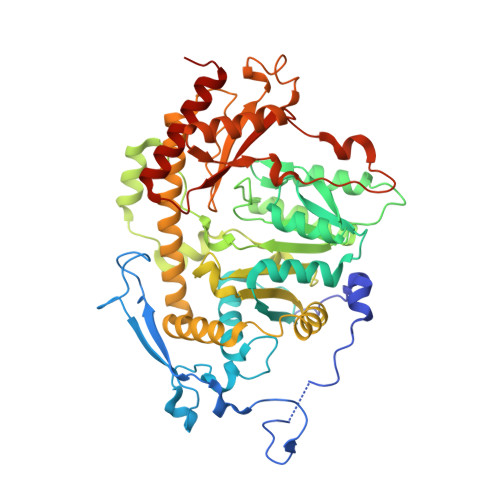
Crystal structure of Saccharomyces cerevisiae Aro8, a putative alpha-aminoadipate aminotransferase.

Bulfer, S.L.Brunzelle, J.S.Trievel, R.C.

(2013) Protein Sci 22: 1417-1424

  • DOI: https://doi.org/10.1002/pro.2315
  • Primary Citation Related Structures: 
    4JE5

  • PubMed Abstract: 

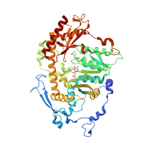
    α-Aminoadipate aminotransferase (AAA-AT) catalyzes the amination of 2-oxoadipate to α-aminoadipate in the fourth step of the α-aminoadipate pathway of lysine biosynthesis in fungi. The aromatic aminotransferase Aro8 has recently been identified as an AAA-AT in Saccharomyces cerevisiae. This enzyme displays broad substrate selectivity, utilizing several amino acids and 2-oxo acids as substrates. Here we report the 1.91Å resolution crystal structure of Aro8 and compare it to AAA-AT LysN from Thermus thermophilus and human kynurenine aminotransferase II. Inspection of the active site of Aro8 reveals asymmetric cofactor binding with lysine-pyridoxal-5-phosphate bound within the active site of one subunit in the Aro8 homodimer and pyridoxamine phosphate and a HEPES molecule bound to the other subunit. The HEPES buffer molecule binds within the substrate-binding site of Aro8, yielding insights into the mechanism by which it recognizes multiple substrates and how this recognition differs from other AAA-AT/kynurenine aminotransferases.


  • Organizational Affiliation
    • Department of Biological Chemistry, University of Michigan, Ann Arbor, Michigan, 48109.

Macromolecule Content 

  • Total Structure Weight: 228.43 kDa 
  • Atom Count: 16,855 
  • Modeled Residue Count: 1,979 
  • Deposited Residue Count: 2,012 
  • Unique protein chains: 2

Macromolecules

Find similar proteins by:|  3D Structure
Entity ID: 1
MoleculeChains  Sequence LengthOrganismDetailsImage
Aromatic/aminoadipate aminotransferase 1
A, B, C
503Saccharomyces cerevisiae S288CMutation(s): 0 
Gene Names: ARO8YGL202W
EC: 2.6.1.39 (PDB Primary Data), 2.6.1.57 (PDB Primary Data)
UniProt
Find proteins for P53090 (Saccharomyces cerevisiae (strain ATCC 204508 / S288c))
Explore P53090 
Go to UniProtKB:  P53090
Entity Groups
Sequence Clusters30% Identity50% Identity70% Identity90% Identity95% Identity100% Identity
UniProt GroupP53090
Sequence Annotations
Expand
Reference Sequence
Find similar proteins by:|  3D Structure
Entity ID: 2
MoleculeChains  Sequence LengthOrganismDetailsImage
Aromatic/aminoadipate aminotransferase 1503Saccharomyces cerevisiae S288CMutation(s): 0 
Gene Names: ARO8YGL202W
EC: 2.6.1.39 (PDB Primary Data), 2.6.1.57 (PDB Primary Data)
UniProt
Find proteins for P53090 (Saccharomyces cerevisiae (strain ATCC 204508 / S288c))
Explore P53090 
Go to UniProtKB:  P53090
Entity Groups
Sequence Clusters30% Identity50% Identity70% Identity90% Identity95% Identity100% Identity
UniProt GroupP53090
Sequence Annotations
Expand
Reference Sequence

Small Molecules

Ligands 3 Unique
IDChains Name / Formula / InChI Key2D Diagram3D Interactions
PMP

Query on PMP


Download:Ideal Coordinates CCD File
I [auth B],
J [auth C]
4'-DEOXY-4'-AMINOPYRIDOXAL-5'-PHOSPHATE
C8 H13 N2 O5 P
ZMJGSOSNSPKHNH-UHFFFAOYSA-N
PLP

Query on PLP


Download:Ideal Coordinates CCD File
E [auth A]PYRIDOXAL-5'-PHOSPHATE
C8 H10 N O6 P
NGVDGCNFYWLIFO-UHFFFAOYSA-N
EPE

Query on EPE


Download:Ideal Coordinates CCD File
F [auth A],
G [auth A],
H [auth B],
K [auth C],
L [auth C]
4-(2-HYDROXYETHYL)-1-PIPERAZINE ETHANESULFONIC ACID
C8 H18 N2 O4 S
JKMHFZQWWAIEOD-UHFFFAOYSA-N
Modified Residues  1 Unique
IDChains TypeFormula2D DiagramParent
LLP
Query on LLP
D
L-PEPTIDE LINKINGC14 H22 N3 O7 PLYS

Experimental Data & Validation

Experimental Data

  • Method: X-RAY DIFFRACTION
  • Resolution: 1.91 Å
  • R-Value Free:  0.217 (Depositor), 0.221 (DCC) 
  • R-Value Work:  0.182 (Depositor), 0.186 (DCC) 
  • R-Value Observed: 0.184 (Depositor) 
Space Group: C 1 2 1
Unit Cell:
Length ( Å )Angle ( ˚ )
a = 227.442α = 90
b = 159.831β = 106.03
c = 70.642γ = 90
Software Package:
Software NamePurpose
DENZOdata reduction
SCALEPACKdata scaling
MOLREPphasing
REFMACrefinement
PDB_EXTRACTdata extraction
HKL-2000data collection

Structure Validation

View Full Validation Report


Ligand Structure Quality Assessment 


Entry History 

Deposition Data

Revision History  (Full details and data files)

  • Version 1.0: 2013-09-11
    Type: Initial release
  • Version 1.1: 2013-10-16
    Changes: Database references
  • Version 1.2: 2013-10-23
    Changes: Database references
  • Version 1.3: 2017-11-15
    Changes: Refinement description
  • Version 1.4: 2025-03-26
    Changes: Data collection, Database references, Derived calculations, Structure summary