4JDJ | pdb_00004jdj

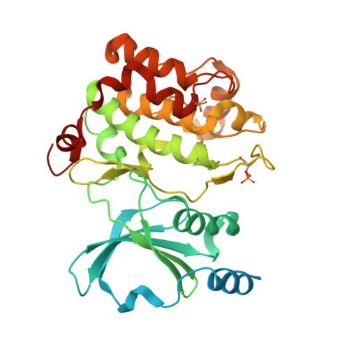
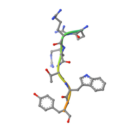
Crystal structure of Serine/threonine-protein kinase PAK 4 F461V mutant in complex with Paktide T peptide substrate


Experimental Data Snapshot

  • Method: X-RAY DIFFRACTION
  • Resolution: 2.30 Å
  • R-Value Free: 
    0.244 (Depositor), 0.215 (DCC) 
  • R-Value Work: 
    0.195 (Depositor) 
  • R-Value Observed: 
    0.197 (Depositor) 

Starting Model: experimental
View more details

wwPDB Validation 3D Report Full Report

Validation slider image for 4JDJ

This is version 1.2 of the entry. See complete history. 

Literature

Identification of a major determinant for serine-threonine kinase phosphoacceptor specificity.

Chen, C., Ha, B.H., Thevenin, A.F., Lou, H.J., Zhang, R., Yip, K.Y., Peterson, J.R., Gerstein, M., Kim, P.M., Filippakopoulos, P., Knapp, S., Boggon, T.J., Turk, B.E.

(2014) Mol Cell 53: 140-147

  • DOI: https://doi.org/10.1016/j.molcel.2013.11.013
  • Primary Citation Related Structures: 
    4JDH, 4JDI, 4JDJ, 4JDK

  • PubMed Abstract: 

    Eukaryotic protein kinases are generally classified as being either tyrosine or serine-threonine specific. Though not evident from inspection of their primary sequences, many serine-threonine kinases display a significant preference for serine or threonine as the phosphoacceptor residue. Here we show that a residue located in the kinase activation segment, which we term the "DFG+1" residue, acts as a major determinant for serine-threonine phosphorylation site specificity. Mutation of this residue was sufficient to switch the phosphorylation site preference for multiple kinases, including the serine-specific kinase PAK4 and the threonine-specific kinase MST4. Kinetic analysis of peptide substrate phosphorylation and crystal structures of PAK4-peptide complexes suggested that phosphoacceptor residue preference is not mediated by stronger binding of the favored substrate. Rather, favored kinase-phosphoacceptor combinations likely promote a conformation optimal for catalysis. Understanding the rules governing kinase phosphoacceptor preference allows kinases to be classified as serine or threonine specific based on their sequence.


  • Organizational Affiliation: 
    • Department of Pharmacology, Yale University School of Medicine, New Haven, CT 06520, USA.

Macromolecule Content 

  • Total Structure Weight: 40.97 kDa 
  • Atom Count: 2,479 
  • Modeled Residue Count: 296 
  • Deposited Residue Count: 361 
  • Unique protein chains: 2

Macromolecules

Find similar proteins by:|  3D Structure
Entity ID: 1
MoleculeChains  Sequence LengthOrganismDetailsImage
Serine/threonine-protein kinase PAK 4346Homo sapiensMutation(s): 1 
Gene Names: KIAA1142, PAK4
EC: 2.7.11.1
UniProt & NIH Common Fund Data Resources
Find proteins for O96013 (Homo sapiens)
Explore O96013 
Go to UniProtKB:  O96013
PHAROS:  O96013
GTEx:  ENSG00000130669 
Entity Groups
Sequence Clusters30% Identity50% Identity70% Identity90% Identity95% Identity100% Identity
UniProt GroupO96013
Sequence Annotations
Expand
Reference Sequence
Find similar proteins by:  Sequence   |   3D Structure  
Entity ID: 2
MoleculeChains  Sequence LengthOrganismDetailsImage
Paktide T15N/AMutation(s): 0 
Entity Groups
Sequence Clusters30% Identity50% Identity70% Identity90% Identity95% Identity100% Identity
Sequence Annotations
Expand
Reference Sequence

Small Molecules

Modified Residues  2 Unique
IDChains TypeFormula2D DiagramParent
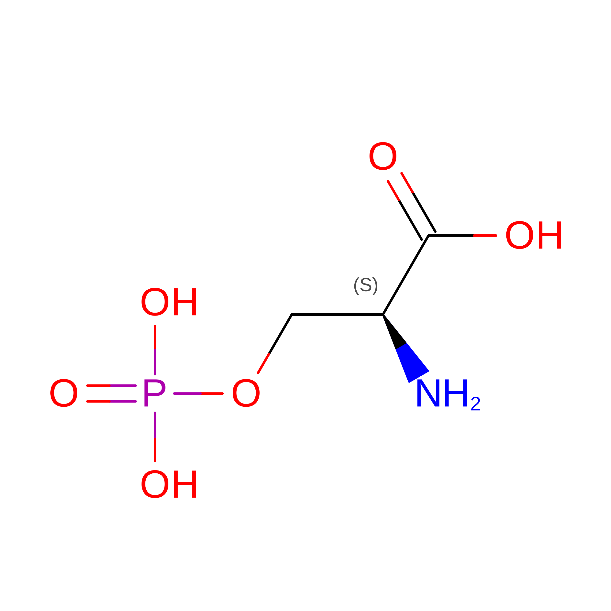
SEP
Query on SEP
A
L-PEPTIDE LINKINGC3 H8 N O6 PSER
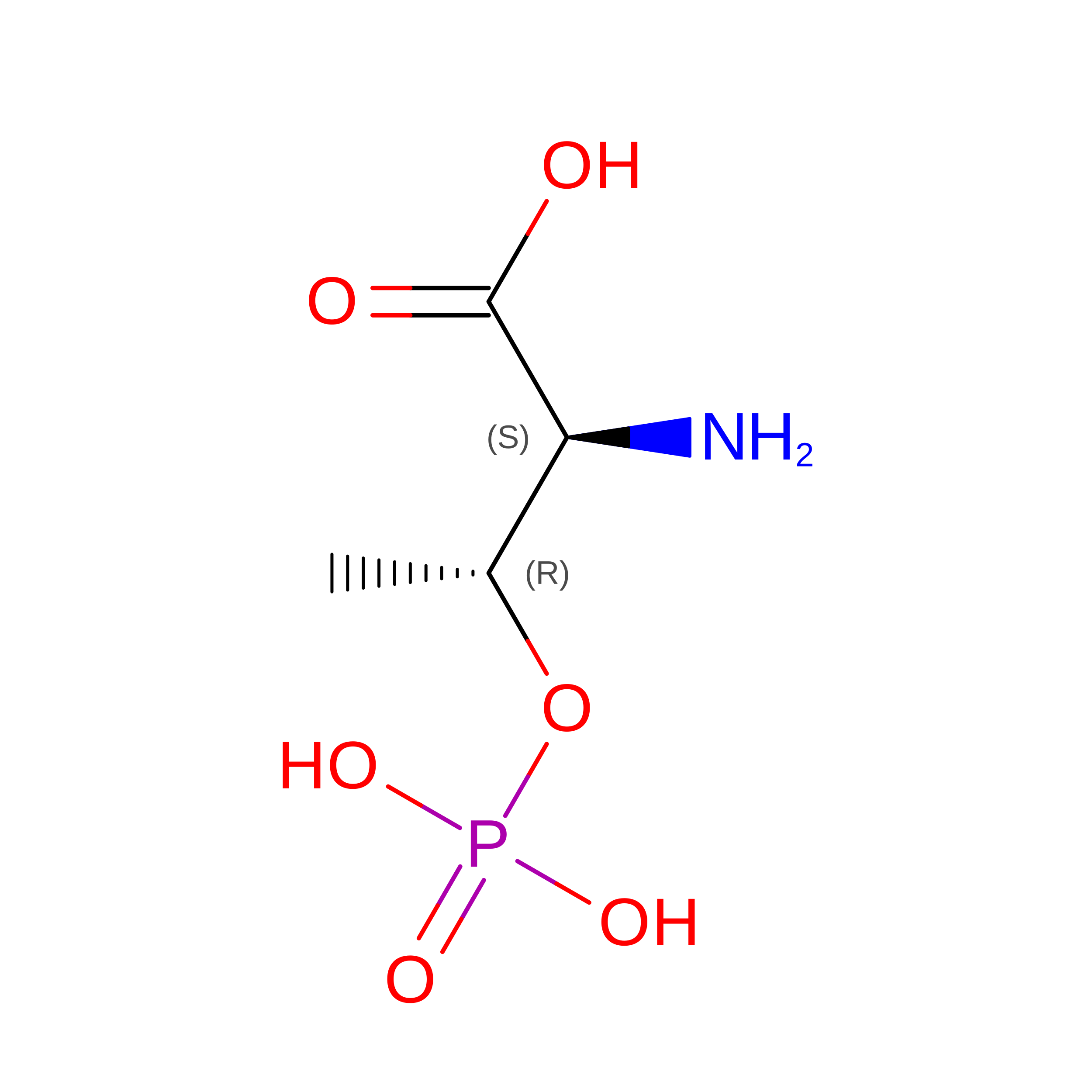
TPO
Query on TPO
A
L-PEPTIDE LINKINGC4 H10 N O6 PTHR

Experimental Data & Validation

Experimental Data

  • Method: X-RAY DIFFRACTION
  • Resolution: 2.30 Å
  • R-Value Free:  0.244 (Depositor), 0.215 (DCC) 
  • R-Value Work:  0.195 (Depositor) 
  • R-Value Observed: 0.197 (Depositor) 
Space Group: P 41 21 2
Unit Cell:
Length ( Å )Angle ( ˚ )
a = 61.79α = 90
b = 61.79β = 90
c = 181.727γ = 90
Software Package:
Software NamePurpose
CBASSdata collection
PHASERphasing
REFMACrefinement
HKL-2000data reduction
HKL-2000data scaling

Structure Validation

View Full Validation Report


Entry History 

Deposition Data

Revision History  (Full details and data files)

  • Version 1.0: 2014-01-29
    Type: Initial release
  • Version 1.1: 2023-09-20
    Changes: Data collection, Database references, Derived calculations, Refinement description
  • Version 1.2: 2024-11-27
    Changes: Structure summary