4J7Q | pdb_00004j7q

Crystal structure of Saccharomyces cerevisiae Sfh3 complexed with phosphatidylinositol


Experimental Data Snapshot

  • Method: X-RAY DIFFRACTION
  • Resolution: 1.55 Å
  • R-Value Free: 
    0.239 (Depositor), 0.238 (DCC) 
  • R-Value Work: 
    0.221 (Depositor), 0.219 (DCC) 
  • R-Value Observed: 
    0.221 (Depositor) 

wwPDB Validation 3D Report Full Report

Validation slider image for 4J7Q

Ligand Structure Quality Assessment 


This is version 1.1 of the entry. See complete history

Literature

Structural determinants for phosphatidylinositol recognition by Sfh3 and substrate-induced dimer-monomer transition during lipid transfer cycles.

Yang, H.Tong, J.Leonard, T.A.Im, Y.J.

(2013) FEBS Lett 587: 1610-1616

  • DOI: https://doi.org/10.1016/j.febslet.2013.04.009
  • Primary Citation Related Structures: 
    4J7P, 4J7Q

  • PubMed Abstract: 

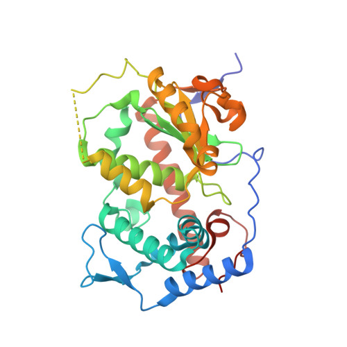
    Sec14 family homologs are the major yeast phosphatidylinositol/phosphatidylcholine transfer proteins regulating lipid metabolism and vesicle trafficking. The structure of Saccharomyces cerevisiae Sfh3 displays a conserved Sec14 scaffold and reveals determinants for the specific recognition of phosphatidylinositol ligand. Apo-Sfh3 forms a dimer through the hydrophobic interaction of gating helices. Binding of phosphatidylinositol leads to dissociation of the dimer into monomers in a reversible manner. This study suggests that the substrate induced dimer-monomer transformation is an essential part of lipid transfer cycles by Sfh3.


  • Organizational Affiliation
    • College of Pharmacy, Chonnam National University, Gwangju 500-757, South Korea.

Macromolecule Content 

  • Total Structure Weight: 78.94 kDa 
  • Atom Count: 5,736 
  • Modeled Residue Count: 644 
  • Deposited Residue Count: 666 
  • Unique protein chains: 1

Macromolecules

Find similar proteins by:|  3D Structure
Entity ID: 1
MoleculeChains  Sequence LengthOrganismDetailsImage
Phosphatidylinositol transfer protein PDR16
A, B
333Saccharomyces cerevisiae S288CMutation(s): 0 
Gene Names: N1158PDR16SFH3YNL231C
UniProt
Find proteins for P53860 (Saccharomyces cerevisiae (strain ATCC 204508 / S288c))
Explore P53860 
Go to UniProtKB:  P53860
Entity Groups
Sequence Clusters30% Identity50% Identity70% Identity90% Identity95% Identity100% Identity
UniProt GroupP53860
Sequence Annotations
Expand
Reference Sequence

Small Molecules

Ligands 1 Unique
IDChains Name / Formula / InChI Key2D Diagram3D Interactions
B7N

Query on B7N


Download:Ideal Coordinates CCD File
C [auth A],
D [auth B]
(1R)-2-{[(S)-hydroxy{[(1S,2R,3R,4S,5S,6R)-2,3,4,5,6-pentahydroxycyclohexyl]oxy}phosphoryl]oxy}-1-[(octadecanoyloxy)methyl]ethyl (9Z)-octadec-9-enoate
C45 H85 O13 P
YOBFISPJJWPPTK-BAAZTTAESA-N

Experimental Data & Validation

Experimental Data

  • Method: X-RAY DIFFRACTION
  • Resolution: 1.55 Å
  • R-Value Free:  0.239 (Depositor), 0.238 (DCC) 
  • R-Value Work:  0.221 (Depositor), 0.219 (DCC) 
  • R-Value Observed: 0.221 (Depositor) 
Space Group: P 1 21 1
Unit Cell:
Length ( Å )Angle ( ˚ )
a = 52.889α = 90
b = 144.149β = 111.28
c = 55.656γ = 90
Software Package:
Software NamePurpose
HKL-2000data collection
MOLREPphasing
CNSrefinement
HKL-2000data reduction
SCALEPACKdata scaling

Structure Validation

View Full Validation Report


Ligand Structure Quality Assessment 


Entry History 

Deposition Data

Revision History  (Full details and data files)

  • Version 1.0: 2013-07-03
    Type: Initial release
  • Version 1.1: 2024-03-20
    Changes: Data collection, Database references, Derived calculations