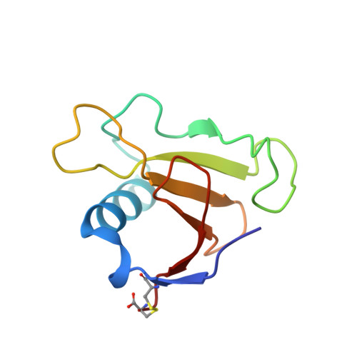
Contribution of hydrogen bonds to protein stability.
Pace, C.N., Fu, H., Lee Fryar, K., Landua, J., Trevino, S.R., Schell, D., Thurlkill, R.L., Imura, S., Scholtz, J.M., Gajiwala, K., Sevcik, J., Urbanikova, L., Myers, J.K., Takano, K., Hebert, E.J., Shirley, B.A., Grimsley, G.R.(2014) Protein Sci 23: 652-661
- PubMed: 24591301
- DOI: https://doi.org/10.1002/pro.2449
- Primary Citation of Related Structures:
4GHO, 4J5G, 4J5K - PubMed Abstract:
Our goal was to gain a better understanding of the contribution of the burial of polar groups and their hydrogen bonds to the conformational stability of proteins. We measured the change in stability, Δ(ΔG), for a series of hydrogen bonding mutants in four proteins: villin headpiece subdomain (VHP) containing 36 residues, a surface protein from Borrelia burgdorferi (VlsE) containing 341 residues, and two proteins previously studied in our laboratory, ribonucleases Sa (RNase Sa) and T1 (RNase T1). Crystal structures were determined for three of the hydrogen bonding mutants of RNase Sa: S24A, Y51F, and T95A. The structures are very similar to wild type RNase Sa and the hydrogen bonding partners form intermolecular hydrogen bonds to water in all three mutants. We compare our results with previous studies of similar mutants in other proteins and reach the following conclusions. (1) Hydrogen bonds contribute favorably to protein stability. (2) The contribution of hydrogen bonds to protein stability is strongly context dependent. (3) Hydrogen bonds by side chains and peptide groups make similar contributions to protein stability. (4) Polar group burial can make a favorable contribution to protein stability even if the polar groups are not hydrogen bonded. (5) The contribution of hydrogen bonds to protein stability is similar for VHP, a small protein, and VlsE, a large protein.
Organizational Affiliation:
Department of Biochemistry and Biophysics, Texas A&M University, College Station, Texas, 77843; Department of Molecular and Cellular Medicine, Texas A&M University Health Science Center, College Station, Texas, 77843.


