4J4B | pdb_00004j4b

PylD in complex with L-lysine-Ne-D-ornithine and NADH


Experimental Data Snapshot

  • Method: X-RAY DIFFRACTION
  • Resolution: 1.90 Å
  • R-Value Free: 
    0.240 (Depositor), 0.240 (DCC) 
  • R-Value Work: 
    0.205 (Depositor), 0.205 (DCC) 
  • R-Value Observed: 
    0.207 (Depositor) 

Starting Model: experimental
View more details

wwPDB Validation 3D Report Full Report

Validation slider image for 4J4B

Ligand Structure Quality Assessment 


This is version 1.3 of the entry. See complete history

Literature

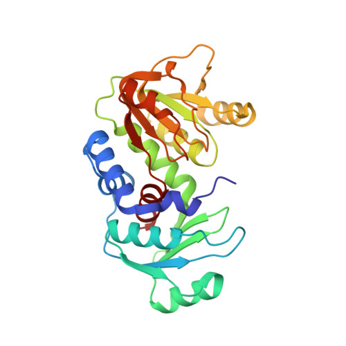
Structure and Reaction Mechanism of Pyrrolysine Synthase (PylD).

Quitterer, F.Beck, P.Bacher, A.Groll, M.

(2013) Angew Chem Int Ed Engl 52: 7033-7037

Macromolecule Content 

  • Total Structure Weight: 57.7 kDa 
  • Atom Count: 4,239 
  • Modeled Residue Count: 518 
  • Deposited Residue Count: 518 
  • Unique protein chains: 1

Macromolecules

Find similar proteins by:|  3D Structure
Entity ID: 1
MoleculeChains  Sequence LengthOrganismDetailsImage
PylD
A, B
259Methanosarcina barkeri str. FusaroMutation(s): 0 
Gene Names: Mbar_A0835
EC: 1.4.1
UniProt
Find proteins for Q46E80 (Methanosarcina barkeri (strain Fusaro / DSM 804))
Explore Q46E80 
Go to UniProtKB:  Q46E80
Entity Groups
Sequence Clusters30% Identity50% Identity70% Identity90% Identity95% Identity100% Identity
UniProt GroupQ46E80
Sequence Annotations
Expand
Reference Sequence

Small Molecules

Ligands 4 Unique
IDChains Name / Formula / InChI Key2D Diagram3D Interactions
NAI

Query on NAI


Download:Ideal Coordinates CCD File
C [auth A],
H [auth B]
1,4-DIHYDRONICOTINAMIDE ADENINE DINUCLEOTIDE
C21 H29 N7 O14 P2
BOPGDPNILDQYTO-NNYOXOHSSA-N
0TF

Query on 0TF


Download:Ideal Coordinates CCD File
F [auth A],
G [auth B]
N~6~-D-ornithyl-L-lysine
C11 H24 N4 O3
AOOVWOOUTNDWMA-BDAKNGLRSA-N
MG

Query on MG


Download:Ideal Coordinates CCD File
E [auth A],
J [auth B]
MAGNESIUM ION
Mg
JLVVSXFLKOJNIY-UHFFFAOYSA-N
NA

Query on NA


Download:Ideal Coordinates CCD File
D [auth A],
I [auth B]
SODIUM ION
Na
FKNQFGJONOIPTF-UHFFFAOYSA-N

Experimental Data & Validation

Experimental Data

  • Method: X-RAY DIFFRACTION
  • Resolution: 1.90 Å
  • R-Value Free:  0.240 (Depositor), 0.240 (DCC) 
  • R-Value Work:  0.205 (Depositor), 0.205 (DCC) 
  • R-Value Observed: 0.207 (Depositor) 
Space Group: C 2 2 21
Unit Cell:
Length ( Å )Angle ( ˚ )
a = 48.383α = 90
b = 142.44β = 90
c = 167.305γ = 90
Software Package:
Software NamePurpose
PROTEUM PLUSdata collection
PHASERphasing
REFMACrefinement
PROTEUM PLUSdata reduction
SCALEPACKdata scaling

Structure Validation

View Full Validation Report


Ligand Structure Quality Assessment 


Entry History 

Deposition Data

Revision History  (Full details and data files)

  • Version 1.0: 2013-06-12
    Type: Initial release
  • Version 1.1: 2013-07-10
    Changes: Database references
  • Version 1.2: 2024-04-03
    Changes: Data collection, Database references, Derived calculations, Refinement description
  • Version 1.3: 2024-10-16
    Changes: Structure summary