4J2T | pdb_00004j2t

Inhibitor-bound Ca2+ ATPase


Experimental Data Snapshot

  • Method: X-RAY DIFFRACTION
  • Resolution: 3.20 Å
  • R-Value Free: 
    0.286 (Depositor), 0.287 (DCC) 
  • R-Value Work: 
    0.260 (Depositor), 0.263 (DCC) 
  • R-Value Observed: 
    0.262 (Depositor) 

Starting Model: experimental
View more details

wwPDB Validation 3D Report Full Report

Validation slider image for 4J2T

Ligand Structure Quality Assessment 


This is version 1.3 of the entry. See complete history

Literature

Water-mediated interactions influence the binding of thapsigargin to sarco/endoplasmic reticulum calcium adenosinetriphosphatase.

Paulsen, E.S.Villadsen, J.Tenori, E.Liu, H.Bonde, D.F.Lie, M.A.Bublitz, M.Olesen, C.Autzen, H.E.Dach, I.Sehgal, P.Nissen, P.Moller, J.V.Schiott, B.Christensen, S.B.

(2013) J Med Chem 56: 3609-3619

  • DOI: https://doi.org/10.1021/jm4001083
  • Primary Citation Related Structures: 
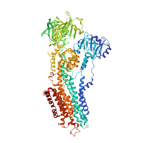
    4J2T

  • PubMed Abstract: 

    A crystal structure suggests four water molecules are present in the binding cavity of thapsigargin in sarco/endoplasmic reticulum calcium ATPase (SERCA). Computational chemistry indicates that three of these water molecules mediate an extensive hydrogen-bonding network between thapsigargin and the backbone of SERCA. The orientation of the thapsigargin molecule in SERCA is crucially dependent on these interactions. The hypothesis has been verified by measuring the affinity of newly synthesized model compounds, which are prevented from participating in such water-mediated interactions as hydrogen-bond donors.


  • Organizational Affiliation
    • Department of Drug Design and Pharmacology, Faculty of Health and Medical Sciences, University of Copenhagen, Universitetsparken 2, DK-2100 Copenhagen Ø, Denmark.

Macromolecule Content 

  • Total Structure Weight: 111.1 kDa 
  • Atom Count: 7,748 
  • Modeled Residue Count: 994 
  • Deposited Residue Count: 994 
  • Unique protein chains: 1

Macromolecules

Find similar proteins by:|  3D Structure
Entity ID: 1
MoleculeChains  Sequence LengthOrganismDetailsImage
SERCA1a994Oryctolagus cuniculusMutation(s): 0 
EC: 3.6.3.8 (PDB Primary Data), 7.2.2.10 (UniProt)
Membrane Entity: Yes 
UniProt
Find proteins for P04191 (Oryctolagus cuniculus)
Explore P04191 
Go to UniProtKB:  P04191
Entity Groups
Sequence Clusters30% Identity50% Identity70% Identity90% Identity95% Identity100% Identity
UniProt GroupP04191
Sequence Annotations
Expand
Reference Sequence

Small Molecules

Ligands 3 Unique
IDChains Name / Formula / InChI Key2D Diagram3D Interactions
PTY

Query on PTY


Download:Ideal Coordinates CCD File
C [auth A]PHOSPHATIDYLETHANOLAMINE
C40 H80 N O8 P
NJGIRBISCGPRPF-KXQOOQHDSA-N
1HT

Query on 1HT


Download:Ideal Coordinates CCD File
B [auth A](3S,3aR,4S,6S,6aR,7S,8S,9bS)-6-(acetyloxy)-3a,4-bis(butanoyloxy)-3-hydroxy-3,6,9-trimethyl-8-{[(2E)-2-methylbut-2-enoyl]oxy}-2-oxo-2,3,3a,4,5,6,6a,7,8,9b-decahydroazuleno[4,5-b]furan-7-yl octanoate
C38 H56 O13
ARWTVHVQANENHV-XDRCOMTCSA-N
K

Query on K


Download:Ideal Coordinates CCD File
D [auth A]POTASSIUM ION
K
NPYPAHLBTDXSSS-UHFFFAOYSA-N

Experimental Data & Validation

Experimental Data

  • Method: X-RAY DIFFRACTION
  • Resolution: 3.20 Å
  • R-Value Free:  0.286 (Depositor), 0.287 (DCC) 
  • R-Value Work:  0.260 (Depositor), 0.263 (DCC) 
  • R-Value Observed: 0.262 (Depositor) 
Space Group: P 41 21 2
Unit Cell:
Length ( Å )Angle ( ˚ )
a = 70.9α = 90
b = 70.9β = 90
c = 587.6γ = 90
Software Package:
Software NamePurpose
MAXLABdata collection
PHASERphasing
PHENIXrefinement
XDSdata reduction
XSCALEdata scaling

Structure Validation

View Full Validation Report


Ligand Structure Quality Assessment 


Entry History 

Revision History  (Full details and data files)

  • Version 1.0: 2013-06-19
    Type: Initial release
  • Version 1.1: 2018-01-24
    Changes: Data collection, Structure summary
  • Version 1.2: 2023-09-20
    Changes: Data collection, Database references, Derived calculations, Refinement description
  • Version 1.3: 2024-10-16
    Changes: Structure summary