4J1Q | pdb_00004j1q

Crystal structure of a ketoreductase domain from the bacillaene assembly line


Experimental Data Snapshot

  • Method: X-RAY DIFFRACTION
  • Resolution: 2.35 Å
  • R-Value Free: 
    0.253 (Depositor), 0.254 (DCC) 
  • R-Value Work: 
    0.232 (Depositor), 0.232 (DCC) 
  • R-Value Observed: 
    0.233 (Depositor) 

wwPDB Validation 3D Report Full Report

Validation slider image for 4J1Q

Ligand Structure Quality Assessment 


This is version 1.4 of the entry. See complete history

Literature

Structural and functional studies of a trans-acyltransferase polyketide assembly line enzyme that catalyzes stereoselective alpha- and beta-ketoreduction.

Piasecki, S.K.Zheng, J.Axelrod, A.J.E Detelich, M.Keatinge-Clay, A.T.

(2014) Proteins 82: 2067-2077

  • DOI: https://doi.org/10.1002/prot.24561
  • Primary Citation Related Structures: 
    4J1Q, 4J1S

  • PubMed Abstract: 

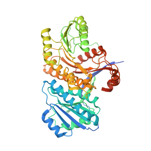
    While the cis-acyltransferase modular polyketide synthase assembly lines have largely been structurally dissected, enzymes from within the recently discovered trans-acyltransferase polyketide synthase assembly lines are just starting to be observed crystallographically. Here we examine the ketoreductase (KR) from the first polyketide synthase module of the bacillaene nonribosomal peptide synthetase/polyketide synthase at 2.35-Å resolution. This KR naturally reduces both α- and β-keto groups and is the only KR known to do so during the biosynthesis of a polyketide. The isolated KR not only reduced an N-acetylcysteamine-bound β-keto substrate to a D-β-hydroxy product, but also an N-acetylcysteamine-bound α-keto substrate to an L-α-hydroxy product. That the substrates must enter the active site from opposite directions to generate these stereochemistries suggests that the acyl-phosphopantetheine moiety is capable of accessing very different conformations despite being anchored to a serine residue of a docked acyl carrier protein. The features enabling stereocontrolled α-ketoreduction may not be extensive since a KR that naturally reduces a β-keto group within a cis-acyltransferase polyketide synthase was identified that performs a completely stereoselective reduction of the same α-keto substrate to generate the D-α-hydroxy product. A sequence analysis of trans-acyltransferase KRs reveals that a single residue, rather than a three-residue motif found in cis-acyltransferase KRs, is predictive of the orientation of the resulting β-hydroxyl group.


  • Organizational Affiliation
    • Institute for Cellular and Molecular Biology, The University of Texas at Austin, Texas, 78712.

Macromolecule Content 

  • Total Structure Weight: 53.26 kDa 
  • Atom Count: 3,378 
  • Modeled Residue Count: 417 
  • Deposited Residue Count: 464 
  • Unique protein chains: 1

Macromolecules

Find similar proteins by:|  3D Structure
Entity ID: 1
MoleculeChains  Sequence LengthOrganismDetailsImage
Polyketide synthase PksJ464Bacillus subtilis subsp. subtilis str. 168Mutation(s): 0 
Gene Names: BSU17180pksJpksJ (amino acids 2669-3111)pksK
EC: 1.1.1.100
UniProt
Find proteins for P40806 (Bacillus subtilis (strain 168))
Explore P40806 
Go to UniProtKB:  P40806
Entity Groups
Sequence Clusters30% Identity50% Identity70% Identity90% Identity95% Identity100% Identity
UniProt GroupP40806
Sequence Annotations
Expand
Reference Sequence

Small Molecules

Ligands 1 Unique
IDChains Name / Formula / InChI Key2D Diagram3D Interactions
NDP

Query on NDP


Download:Ideal Coordinates CCD File
B [auth A]NADPH DIHYDRO-NICOTINAMIDE-ADENINE-DINUCLEOTIDE PHOSPHATE
C21 H30 N7 O17 P3
ACFIXJIJDZMPPO-NNYOXOHSSA-N

Experimental Data & Validation

Experimental Data

  • Method: X-RAY DIFFRACTION
  • Resolution: 2.35 Å
  • R-Value Free:  0.253 (Depositor), 0.254 (DCC) 
  • R-Value Work:  0.232 (Depositor), 0.232 (DCC) 
  • R-Value Observed: 0.233 (Depositor) 
Space Group: P 61
Unit Cell:
Length ( Å )Angle ( ˚ )
a = 59.529α = 90
b = 59.529β = 90
c = 220.792γ = 120
Software Package:
Software NamePurpose
HKL-2000data collection
PHENIXmodel building
REFMACrefinement
HKL-2000data reduction
HKL-2000data scaling
PHENIXphasing

Structure Validation

View Full Validation Report


Ligand Structure Quality Assessment 


Entry History 

Deposition Data

Revision History  (Full details and data files)

  • Version 1.0: 2014-03-19
    Type: Initial release
  • Version 1.1: 2014-03-26
    Changes: Database references
  • Version 1.2: 2014-08-13
    Changes: Database references
  • Version 1.3: 2014-09-03
    Changes: Database references
  • Version 1.4: 2024-02-28
    Changes: Data collection, Database references, Derived calculations