4IWJ | pdb_00004iwj

Crystal structure of iron soaked (45 min) ferritin from Pseudo-nitzschia multiseries


Experimental Data Snapshot

  • Method: X-RAY DIFFRACTION
  • Resolution: 1.95 Å
  • R-Value Free: 
    0.213 (Depositor), 0.213 (DCC) 
  • R-Value Work: 
    0.173 (Depositor), 0.173 (DCC) 
  • R-Value Observed: 
    0.175 (Depositor) 

wwPDB Validation 3D Report Full Report

Validation slider image for 4IWJ

This is version 1.3 of the entry. See complete history

Literature

Mechanism of ferrous iron binding and oxidation by ferritin from a pennate diatom.

Pfaffen, S.Abdulqadir, R.Le Brun, N.E.Murphy, M.E.

(2013) J Biological Chem 288: 14917-14925

  • DOI: https://doi.org/10.1074/jbc.M113.454496
  • Primary Citation Related Structures: 
    4ISM, 4ISP, 4ITT, 4ITW, 4IWJ, 4IWK, 4IXK

  • PubMed Abstract: 

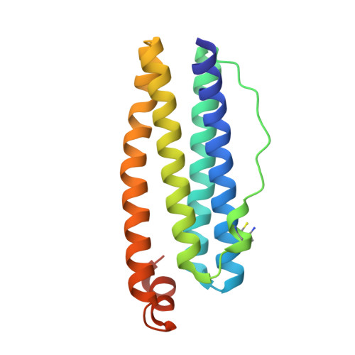
    A novel ferritin was recently found in Pseudo-nitzschia multiseries (PmFTN), a marine pennate diatom that plays a major role in global primary production and carbon sequestration into the deep ocean. Crystals of recombinant PmFTN were soaked in iron and zinc solutions, and the structures were solved to 1.65-2.2-Å resolution. Three distinct iron binding sites were identified as determined from anomalous dispersion data from aerobically grown ferrous soaked crystals. Sites A and B comprise the conserved ferroxidase active site, and site C forms a pathway leading toward the central cavity where iron storage occurs. In contrast, crystal structures derived from anaerobically grown and ferrous soaked crystals revealed only one ferrous iron in the active site occupying site A. In the presence of dioxygen, zinc is observed bound to all three sites. Iron oxidation experiments using stopped-flow absorbance spectroscopy revealed an extremely rapid phase corresponding to Fe(II) oxidation at the ferroxidase site, which is saturated after adding 48 ferrous iron to apo-PmFTN (two ferrous iron per subunit), and a much slower phase due to iron core formation. These results suggest an ordered stepwise binding of ferrous iron and dioxygen to the ferroxidase site in preparation for catalysis and a partial mobilization of iron from the site following oxidation.


  • Organizational Affiliation
    • Department of Microbiology and Immunology, University of British Columbia, Vancouver, British Columbia V6T 1Z3, Canada.

Macromolecule Content 

  • Total Structure Weight: 151.83 kDa 
  • Atom Count: 11,333 
  • Modeled Residue Count: 1,269 
  • Deposited Residue Count: 1,344 
  • Unique protein chains: 1

Macromolecules

Find similar proteins by:|  3D Structure
Entity ID: 1
MoleculeChains  Sequence LengthOrganismDetailsImage
Ferritin168Pseudo-nitzschia multiseriesMutation(s): 0 
Gene Names: FTN
EC: 1.16.3.1
UniProt
Find proteins for B6DMH6 (Pseudo-nitzschia multiseries)
Explore B6DMH6 
Go to UniProtKB:  B6DMH6
Entity Groups
Sequence Clusters30% Identity50% Identity70% Identity90% Identity95% Identity100% Identity
UniProt GroupB6DMH6
Sequence Annotations
Expand
Reference Sequence

Small Molecules

Ligands 1 Unique
IDChains Name / Formula / InChI Key2D Diagram3D Interactions
FE

Query on FE


Download:Ideal Coordinates CCD File
I [auth A]
J [auth A]
K [auth D]
L [auth D]
M [auth B]
I [auth A],
J [auth A],
K [auth D],
L [auth D],
M [auth B],
N [auth B],
O [auth C],
P [auth C],
Q [auth E],
R [auth E],
S [auth F],
T [auth F],
U [auth G],
V [auth G],
W [auth H],
X [auth H]
FE (III) ION
Fe
VTLYFUHAOXGGBS-UHFFFAOYSA-N
Modified Residues  1 Unique
IDChains TypeFormula2D DiagramParent
YCM
Query on YCM
A
B [auth D]
C [auth B]
D [auth C]
E
A,
B [auth D],
C [auth B],
D [auth C],
E,
F,
G,
H
L-PEPTIDE LINKINGC5 H10 N2 O3 SCYS

Experimental Data & Validation

Experimental Data

  • Method: X-RAY DIFFRACTION
  • Resolution: 1.95 Å
  • R-Value Free:  0.213 (Depositor), 0.213 (DCC) 
  • R-Value Work:  0.173 (Depositor), 0.173 (DCC) 
  • R-Value Observed: 0.175 (Depositor) 
Space Group: P 2 3
Unit Cell:
Length ( Å )Angle ( ˚ )
a = 175.319α = 90
b = 175.319β = 90
c = 175.319γ = 90
Software Package:
Software NamePurpose
DENZOdata reduction
SCALEPACKdata scaling
MOLREPphasing
REFMACrefinement
PDB_EXTRACTdata extraction

Structure Validation

View Full Validation Report


Entry History 

Deposition Data

Revision History  (Full details and data files)

  • Version 1.0: 2013-03-20
    Type: Initial release
  • Version 1.1: 2013-06-19
    Changes: Database references
  • Version 1.2: 2017-11-15
    Changes: Refinement description
  • Version 1.3: 2025-03-26
    Changes: Data collection, Database references, Derived calculations, Structure summary