4IUJ | pdb_00004iuj

Structure of Polymerase acid protein (PA) from Influenzavirus A Influenza A virus A, WILSON-SMITH/1933 (H1N1)


Experimental Data Snapshot

  • Method: X-RAY DIFFRACTION
  • Resolution: 1.90 Å
  • R-Value Free: 
    0.201 (Depositor), 0.208 (DCC) 
  • R-Value Work: 
    0.174 (Depositor), 0.184 (DCC) 
  • R-Value Observed: 
    0.175 (Depositor) 

Starting Model: experimental
View more details

wwPDB Validation 3D Report Full Report

Validation slider image for 4IUJ

This is version 1.3 of the entry. See complete history

Literature

Structural analysis of H1N1 and H7N9 influenza A virus PA in the absence of PB1.

Moen, S.O.Abendroth, J.Fairman, J.W.Baydo, R.O.Bullen, J.Kirkwood, J.L.Barnes, S.R.Raymond, A.C.Begley, D.W.Henkel, G.McCormack, K.Tam, V.C.Phan, I.Staker, B.L.Stacy, R.Myler, P.J.Lorimer, D.Edwards, T.E.

(2014) Sci Rep 4: 5944-5944

  • DOI: https://doi.org/10.1038/srep05944
  • Primary Citation Related Structures: 
    4IUJ, 4P9A

  • PubMed Abstract: 

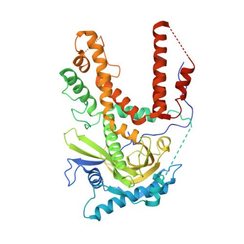
    Influenza A viruses cause the respiratory illness influenza, which can be mild to fatal depending on the strain and host immune response. The flu polymerase acidic (PA), polymerase basic 1 (PB1), and polymerase basic 2 (PB2) proteins comprise the RNA-dependent RNA polymerase complex responsible for viral genome replication. The first crystal structures of the C-terminal domain of PA (PA-CTD) in the absence of PB1-derived peptides show a number of structural changes relative to the previously reported PB1-peptide bound structures. The human A/WSN/1933 (H1N1) and avian A/Anhui1/2013 (H7N9) strain PA-CTD proteins exhibit the same global topology as other strains in the absence of PB1, but differ extensively in the PB1 binding pocket including a widening of the binding groove and the unfolding of a β-turn. Both PA-CTD proteins exhibited a significant increase in thermal stability in the presence of either a PB1-derived peptide or a previously reported inhibitor in differential scanning fluorimetry assays. These structural changes demonstrate plasticity in the PA-PB1 binding interface which may be exploited in the development of novel therapeutics.


  • Organizational Affiliation
    • 1] Seattle Structural Genomics Center for Infectious Disease (SSGCID) [2] Beryllium, 7869 NE Day Road West, Bainbridge Island, WA 98110, USA.

Macromolecule Content 

  • Total Structure Weight: 53.1 kDa 
  • Atom Count: 3,484 
  • Modeled Residue Count: 408 
  • Deposited Residue Count: 464 
  • Unique protein chains: 1

Macromolecules

Find similar proteins by:|  3D Structure
Entity ID: 1
MoleculeChains  Sequence LengthOrganismDetailsImage
Polymerase acidic protein464Influenza A virus (A/Wilson-Smith/1933(H1N1))Mutation(s): 0 
Gene Names: PA
EC: 3.1
UniProt
Find proteins for P15659 (Influenza A virus (strain A/Wilson-Smith/1933 H1N1))
Explore P15659 
Go to UniProtKB:  P15659
Entity Groups
Sequence Clusters30% Identity50% Identity70% Identity90% Identity95% Identity100% Identity
UniProt GroupP15659
Sequence Annotations
Expand
Reference Sequence

Experimental Data & Validation

Experimental Data

  • Method: X-RAY DIFFRACTION
  • Resolution: 1.90 Å
  • R-Value Free:  0.201 (Depositor), 0.208 (DCC) 
  • R-Value Work:  0.174 (Depositor), 0.184 (DCC) 
  • R-Value Observed: 0.175 (Depositor) 
Space Group: P 65 2 2
Unit Cell:
Length ( Å )Angle ( ˚ )
a = 68.8α = 90
b = 68.8β = 90
c = 395.65γ = 120
Software Package:
Software NamePurpose
XSCALEdata scaling
PHASERphasing
REFMACrefinement
PDB_EXTRACTdata extraction
XDSdata reduction

Structure Validation

View Full Validation Report


Entry History 

Deposition Data

Revision History  (Full details and data files)

  • Version 1.0: 2013-02-06
    Type: Initial release
  • Version 1.1: 2014-08-20
    Changes: Database references
  • Version 1.2: 2017-11-15
    Changes: Refinement description
  • Version 1.3: 2023-09-20
    Changes: Data collection, Database references, Refinement description