4ITR | pdb_00004itr

Crystal Structure of IbpAFic2-H3717A in complex with adenylylated Cdc42


Experimental Data Snapshot

  • Method: X-RAY DIFFRACTION
  • Resolution: 2.30 Å
  • R-Value Free: 
    0.226 (Depositor), 0.204 (DCC) 
  • R-Value Work: 
    0.178 (Depositor) 
  • R-Value Observed: 
    0.181 (Depositor) 

wwPDB Validation 3D Report Full Report

Validation slider image for 4ITR

Ligand Structure Quality Assessment 


This is version 3.0 of the entry. See complete history

Literature

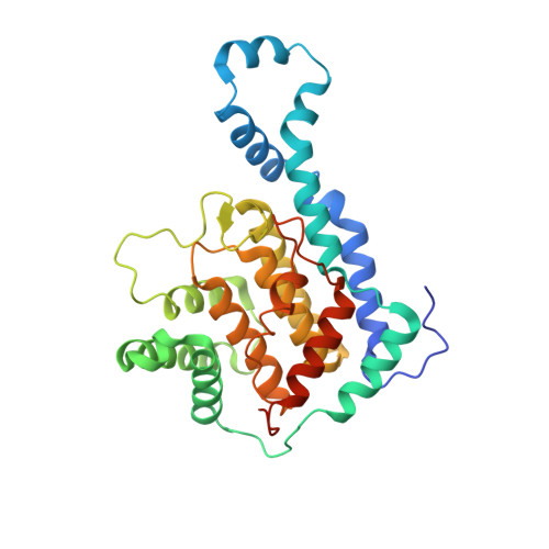
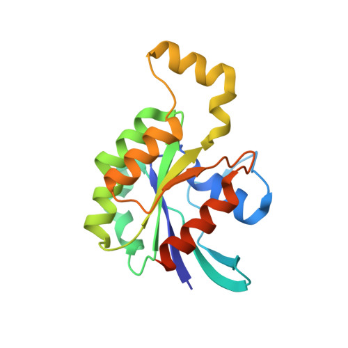
Structural basis of Fic-mediated adenylylation.

Xiao, J.Worby, C.A.Mattoo, S.Sankaran, B.Dixon, J.E.

(2010) Nat Struct Mol Biol 17: 1004-1010

  • DOI: https://doi.org/10.1038/nsmb.1867
  • Primary Citation Related Structures: 
    3N3U, 4ITR

  • PubMed Abstract: 

    The Fic family of adenylyltransferases, defined by a core HPFx(D/E)GN(G/K)R motif, consists of over 2,700 proteins found in organisms from bacteria to humans. The immunoglobulin-binding protein A (IbpA) from the bacterial pathogen Histophilus somni contains two Fic domains that adenylylate the switch1 tyrosine residue of Rho-family GTPases, allowing the bacteria to subvert host defenses. Here we present the structure of the second Fic domain of IbpA (IbpAFic2) in complex with its substrate, Cdc42. IbpAFic2-bound Cdc42 mimics the GDI-bound state of Rho GTPases, with both its switch1 and switch2 regions gripped by IbpAFic2. Mutations disrupting the IbpAFic2-Cdc42 interface impair adenylylation and cytotoxicity. Notably, the switch1 tyrosine of Cdc42 is adenylylated in the structure, providing the first structural view for this post-translational modification. We also show that the nucleotide-binding mechanism is conserved among Fic proteins and propose a catalytic mechanism for this recently discovered family of enzymes.


  • Organizational Affiliation
    • Department of Pharmacology, Howard Hughes Medical Institute, University of California, San Diego, La Jolla, California, USA.

Macromolecule Content 

  • Total Structure Weight: 115.57 kDa 
  • Atom Count: 8,384 
  • Modeled Residue Count: 948 
  • Deposited Residue Count: 1,014 
  • Unique protein chains: 2

Macromolecules

Find similar proteins by:|  3D Structure
Entity ID: 1
MoleculeChains  Sequence LengthOrganismDetailsImage
Adenosine monophosphate-protein transferase and cysteine protease IbpA
A, B
316Histophilus somni 2336Mutation(s): 1 
Gene Names: ibpAp76HSM_1489
EC: 2.7.7.1 (PDB Primary Data), 3.4.22 (UniProt), 2.7.7.108 (UniProt)
UniProt
Find proteins for Q06277 (Histophilus somni (strain 2336))
Explore Q06277 
Go to UniProtKB:  Q06277
Entity Groups
Sequence Clusters30% Identity50% Identity70% Identity90% Identity95% Identity100% Identity
UniProt GroupQ06277
Sequence Annotations
Expand
Reference Sequence
Find similar proteins by:|  3D Structure
Entity ID: 2
MoleculeChains  Sequence LengthOrganismDetailsImage
Cell division control protein 42 homolog
C, D
191Homo sapiensMutation(s): 0 
Gene Names: CDC42
EC: 3.6.5.2
UniProt & NIH Common Fund Data Resources
Find proteins for P60953 (Homo sapiens)
Explore P60953 
Go to UniProtKB:  P60953
PHAROS:  P60953
GTEx:  ENSG00000070831 
Entity Groups
Sequence Clusters30% Identity50% Identity70% Identity90% Identity95% Identity100% Identity
UniProt GroupP60953
Sequence Annotations
Expand
Reference Sequence

Small Molecules

Ligands 4 Unique
IDChains Name / Formula / InChI Key2D Diagram3D Interactions
GDP

Query on GDP


Download:Ideal Coordinates CCD File
H [auth C],
L [auth D]
GUANOSINE-5'-DIPHOSPHATE
C10 H15 N5 O11 P2
QGWNDRXFNXRZMB-UUOKFMHZSA-N
AMP

Query on AMP


Download:Ideal Coordinates CCD File
G [auth C],
K [auth D]
ADENOSINE MONOPHOSPHATE
C10 H14 N5 O7 P
UDMBCSSLTHHNCD-KQYNXXCUSA-N
SO4

Query on SO4


Download:Ideal Coordinates CCD File
E [auth A],
F [auth B],
J [auth C],
N [auth D]
SULFATE ION
O4 S
QAOWNCQODCNURD-UHFFFAOYSA-L
MG

Query on MG


Download:Ideal Coordinates CCD File
I [auth C],
M [auth D]
MAGNESIUM ION
Mg
JLVVSXFLKOJNIY-UHFFFAOYSA-N

Experimental Data & Validation

Experimental Data

  • Method: X-RAY DIFFRACTION
  • Resolution: 2.30 Å
  • R-Value Free:  0.226 (Depositor), 0.204 (DCC) 
  • R-Value Work:  0.178 (Depositor) 
  • R-Value Observed: 0.181 (Depositor) 
Space Group: P 21 21 21
Unit Cell:
Length ( Å )Angle ( ˚ )
a = 63.495α = 90
b = 90.997β = 90
c = 190.969γ = 90
Software Package:
Software NamePurpose
PHENIXrefinement

Structure Validation

View Full Validation Report


Ligand Structure Quality Assessment 


Entry History 

Deposition Data

Revision History  (Full details and data files)

  • Version 1.0: 2013-02-20
    Type: Initial release
  • Version 2.0: 2023-06-07
    Changes: Advisory, Atomic model, Data collection, Database references, Derived calculations
  • Version 2.1: 2024-11-06
    Changes: Data collection, Structure summary
  • Version 3.0: 2025-04-23
    Changes: Atomic model