4IRP | pdb_00004irp

Crystal structure of catalytic domain of human beta1,4-galactosyltransferase-7 in open conformation with manganses and UDP


Experimental Data Snapshot

  • Method: X-RAY DIFFRACTION
  • Resolution: 2.10 Å
  • R-Value Free: 
    0.275 (Depositor), 0.276 (DCC) 
  • R-Value Work: 
    0.218 (Depositor), 0.221 (DCC) 
  • R-Value Observed: 
    0.221 (Depositor) 

Starting Model: experimental
View more details

wwPDB Validation 3D Report Full Report

Validation slider image for 4IRP

Ligand Structure Quality Assessment 


This is version 1.3 of the entry. See complete history

Literature

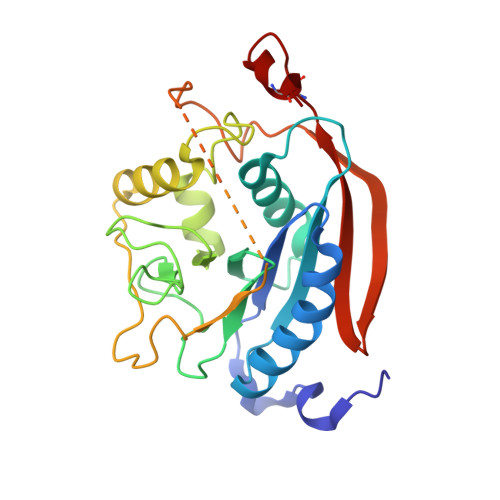
Crystal Structures of beta-1,4-Galactosyltransferase 7 Enzyme Reveal Conformational Changes and Substrate Binding.

Tsutsui, Y.Ramakrishnan, B.Qasba, P.K.

(2013) J Biological Chem 288: 31963-31970

  • DOI: https://doi.org/10.1074/jbc.M113.509984
  • Primary Citation Related Structures: 
    4IRP, 4IRQ, 4LW3, 4LW6, 4M4K

  • PubMed Abstract: 

    The β-1,4-galactosyltransferase 7 (β4GalT7) enzyme is involved in proteoglycan synthesis. In the presence of a manganese ion, it transfers galactose from UDP-galactose to xylose on a proteoglycan acceptor substrate. We present here the crystal structures of human β4GalT7 in open and closed conformations. A comparison of these crystal structures shows that, upon manganese and UDP or UDP-Gal binding, the enzyme undergoes conformational changes involving a small and a long loop. We also present the crystal structures of Drosophila wild-type β4GalT7 and D211N β4GalT7 mutant enzymes in the closed conformation in the presence of the acceptor substrate xylobiose and the donor substrate UDP-Gal, respectively. To understand the catalytic mechanism, we have crystallized the ternary complex of D211N β4GalT7 mutant enzyme in the presence of manganese with the donor and the acceptor substrates together in the same crystal structure. The galactose moiety of the bound UDP-Gal molecule forms seven hydrogen bonds with the protein molecule. The nonreducing end of the xylose moiety of xylobiose binds to the hydrophobic acceptor sugar binding pocket created by the conformational changes, whereas its extended xylose moiety forms hydrophobic interactions with a Tyr residue. In the ternary complex crystal structure, the nucleophile O4 oxygen atom of the xylose molecule is found in close proximity to the C1 and O5 atoms of the galactose moiety. This is the first time that a Michaelis complex of a glycosyltransferase has been described, and it clearly suggests an SN2 type catalytic mechanism for the β4GalT7 enzyme.


  • Organizational Affiliation
    • From the Structural Glycobiology Section and Basic Research Program, SAIC-Frederick, Inc., Nanobiology Program, Center for Cancer Research, NCI, National Institutes of Health, Frederick, Maryland 21702.

Macromolecule Content 

  • Total Structure Weight: 59.77 kDa 
  • Atom Count: 4,288 
  • Modeled Residue Count: 459 
  • Deposited Residue Count: 502 
  • Unique protein chains: 1

Macromolecules

Find similar proteins by:|  3D Structure
Entity ID: 1
MoleculeChains  Sequence LengthOrganismDetailsImage
Beta-1,4-galactosyltransferase 7
A, B
251Homo sapiensMutation(s): 0 
Gene Names: 4galactosyltransferaseB4GALT7beta1UNQ748/PRO1478XGALT1
EC: 2.4.1 (PDB Primary Data), 2.4.1.133 (PDB Primary Data)
UniProt & NIH Common Fund Data Resources
Find proteins for Q9UBV7 (Homo sapiens)
Explore Q9UBV7 
Go to UniProtKB:  Q9UBV7
PHAROS:  Q9UBV7
GTEx:  ENSG00000027847 
Entity Groups
Sequence Clusters30% Identity50% Identity70% Identity90% Identity95% Identity100% Identity
UniProt GroupQ9UBV7
Sequence Annotations
Expand
Reference Sequence

Small Molecules

Ligands 4 Unique
IDChains Name / Formula / InChI Key2D Diagram3D Interactions
UDP

Query on UDP


Download:Ideal Coordinates CCD File
C [auth A],
J [auth B]
URIDINE-5'-DIPHOSPHATE
C9 H14 N2 O12 P2
XCCTYIAWTASOJW-XVFCMESISA-N
IMD

Query on IMD


Download:Ideal Coordinates CCD File
I [auth A]IMIDAZOLE
C3 H5 N2
RAXXELZNTBOGNW-UHFFFAOYSA-O
ACT

Query on ACT


Download:Ideal Coordinates CCD File
E [auth A]
F [auth A]
G [auth A]
H [auth A]
L [auth B]
E [auth A],
F [auth A],
G [auth A],
H [auth A],
L [auth B],
M [auth B]
ACETATE ION
C2 H3 O2
QTBSBXVTEAMEQO-UHFFFAOYSA-M
MN

Query on MN


Download:Ideal Coordinates CCD File
D [auth A],
K [auth B]
MANGANESE (II) ION
Mn
WAEMQWOKJMHJLA-UHFFFAOYSA-N

Experimental Data & Validation

Experimental Data

  • Method: X-RAY DIFFRACTION
  • Resolution: 2.10 Å
  • R-Value Free:  0.275 (Depositor), 0.276 (DCC) 
  • R-Value Work:  0.218 (Depositor), 0.221 (DCC) 
  • R-Value Observed: 0.221 (Depositor) 
Space Group: P 41 21 2
Unit Cell:
Length ( Å )Angle ( ˚ )
a = 125.186α = 90
b = 125.186β = 90
c = 87.326γ = 90
Software Package:
Software NamePurpose
HKL-2000data collection
PHENIXmodel building
PHENIXrefinement
HKL-2000data reduction
HKL-2000data scaling
PHENIXphasing

Structure Validation

View Full Validation Report


Ligand Structure Quality Assessment 


Entry History 

Deposition Data

Revision History  (Full details and data files)

  • Version 1.0: 2013-09-25
    Type: Initial release
  • Version 1.1: 2013-11-27
    Changes: Database references
  • Version 1.2: 2023-09-20
    Changes: Data collection, Database references, Derived calculations, Refinement description
  • Version 1.3: 2024-11-27
    Changes: Structure summary