Structure of the prolyl-acyl carrier protein oxidase involved in the biosynthesis of the cyanotoxin anatoxin-a.
Moncoq, K., Regad, L., Mann, S., Mejean, A., Ploux, O.(2013) Acta Crystallogr D Biol Crystallogr 69: 2340-2352
- PubMed: 24311576 Search on PubMed
- DOI: https://doi.org/10.1107/S0907444913021859
- Primary Citation Related Structures:
4IRN - PubMed Abstract:
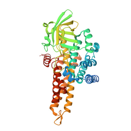
Anatoxin-a and homoanatoxin-a are two potent cyanobacterial neurotoxins biosynthesized from L-proline by a short pathway involving polyketide synthases. Proline is first loaded onto AnaD, an acyl carrier protein, and prolyl-AnaD is then oxidized to 1-pyrroline-5-carboxyl-AnaD by a flavoprotein, AnaB. Three polyketide synthases then transform this imine into anatoxin-a or homoanatoxin-a. AnaB was crystallized in its holo form and its three-dimensional structure was determined by X-ray diffraction at 2.8 Å resolution. AnaB is a homotetramer and its fold is very similar to that of the acyl-CoA dehydrogenases (ACADs). The active-site base of AnaB, Glu244, superimposed very well with that of human isovaleryl-CoA dehydrogenase, confirming previous site-directed mutagenesis experiments and mechanistic proposals. The substrate-binding site of AnaB is small and is likely to be fitted for the pyrrolidine ring of proline. However, in contrast to ACADs, which use an electron-transport protein, AnaB uses molecular oxygen as the electron acceptor, as in acyl-CoA oxidases. Calculation of the solvent-accessible surface area around the FAD in AnaB and in several homologues showed that it is significantly larger in AnaB than in its homologues. A protonated histidine near the FAD in AnaB is likely to participate in oxygen activation. Furthermore, an array of water molecules detected in the AnaB structure suggests a possible path for molecular oxygen towards FAD. This is consistent with AnaB being an oxidase rather than a dehydrogenase. The structure of AnaB is the first to be described for a prolyl-ACP oxidase and it will contribute to defining the structural basis responsible for oxygen reactivity in flavoenzymes.
- CNRS, UMR 7099, Institut de Biologie Physico-Chimique, 13 Rue Pierre et Marie Curie, 75005 Paris, France.
Organizational Affiliation: