4IOV | pdb_00004iov

The structure of AAVrh32.33, a Novel Gene Delivery Vector


Experimental Data Snapshot

  • Method: X-RAY DIFFRACTION
  • Resolution: 3.50 Å
  • R-Value Free: 
    0.269 (Depositor), 0.270 (DCC) 
  • R-Value Work: 
    0.271 (Depositor), 0.269 (DCC) 
  • R-Value Observed: 
    0.271 (Depositor) 

wwPDB Validation 3D Report Full Report

Validation slider image for 4IOV

Ligand Structure Quality Assessment 


This is version 1.2 of the entry. See complete history

Literature

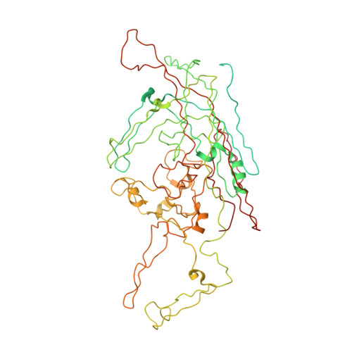
The structure of AAVrh32.33, a novel gene delivery vector.

Mikals, K.Nam, H.J.Van Vliet, K.Vandenberghe, L.H.Mays, L.E.McKenna, R.Wilson, J.M.Agbandje-McKenna, M.

(2014) J Struct Biol 186: 308-317

  • DOI: https://doi.org/10.1016/j.jsb.2014.03.020
  • Primary Citation Related Structures: 
    4IOV

  • PubMed Abstract: 

    The Adeno-associated viruses (AAVs) are being developed as gene delivery vectors for therapeutic clinical applications. However, the host antibody immune response directed against their capsid, prevalent in ∼40-70% of the general population, depending on serotype, negatively impacts efficacy. AAVrh32.33, a novel vector developed from rhesus macaques isolates, has significantly lower seroprevalence in human populations compared to AAV2 and AAV8, which are both in clinical use. To better understand the capsid determinants of this differential immune response to AAVrh32.33, its structure was determined by X-ray crystallography to 3.5 Å resolution. The capsid viral protein (VP) structure conserves the eight-stranded β-barrel core and αA helix reported for other parvoviruses and the distinct capsid surface topology of the AAVs: a depression at the icosahedral twofold axis, three protrusions surrounding the threefold axis, and a depression surround a cylindrical channel at the fivefold axis. A comparison to AAV2, AAV4, and AAV8, to which AAVrh32.33 shares ∼61%, ∼81%, and ∼63% identity, respectively, identified differences in previously defined AAV VP structurally variable regions (VR-1 to VR-IX) which function as receptor attachment, transduction efficiency, and/or antigenic determinants. This structure thus provides a 3D platform for capsid engineering in ongoing efforts to develop AAVrh32.33, as well as other AAV serotypes, for tissue targeted gene-therapy applications with vectors that can evade pre-existing antibody responses against the capsid. These features are required for full clinical realization of the promising AAV gene delivery system.


  • Organizational Affiliation
    • Department of Biochemistry and Molecular Biology, Center for Structural Biology, The McKnight Brain Institute, University of Florida, Gainesville, FL, USA.

Macromolecule Content 

  • Total Structure Weight: 81.49 kDa 
  • Atom Count: 4,139 
  • Modeled Residue Count: 520 
  • Deposited Residue Count: 733 
  • Unique protein chains: 1

Macromolecules

Find similar proteins by:|  3D Structure
Entity ID: 1
MoleculeChains  Sequence LengthOrganismDetailsImage
Capsid protein VP1733Adeno-associated virusMutation(s): 0 
Gene Names: Adeno associated virus Rh32-33 VP1
UniProt
Find proteins for B4Y891 (Adeno-associated virus)
Explore B4Y891 
Go to UniProtKB:  B4Y891
Entity Groups
Sequence Clusters30% Identity50% Identity70% Identity90% Identity95% Identity100% Identity
UniProt GroupB4Y891
Sequence Annotations
Expand
Reference Sequence

Small Molecules

Ligands 1 Unique
IDChains Name / Formula / InChI Key2D Diagram3D Interactions
D5M

Query on D5M


Download:Ideal Coordinates CCD File
B [auth A]2'-DEOXYADENOSINE-5'-MONOPHOSPHATE
C10 H14 N5 O6 P
KHWCHTKSEGGWEX-RRKCRQDMSA-N

Experimental Data & Validation

Experimental Data

  • Method: X-RAY DIFFRACTION
  • Resolution: 3.50 Å
  • R-Value Free:  0.269 (Depositor), 0.270 (DCC) 
  • R-Value Work:  0.271 (Depositor), 0.269 (DCC) 
  • R-Value Observed: 0.271 (Depositor) 
Space Group: P 1
Unit Cell:
Length ( Å )Angle ( ˚ )
a = 246.396α = 70.38
b = 247.521β = 65.37
c = 250.332γ = 60.17
Software Package:
Software NamePurpose
HKL-2000data collection
AMoREphasing
CNSrefinement
HKL-2000data reduction
HKL-2000data scaling

Structure Validation

View Full Validation Report


Ligand Structure Quality Assessment 


Entry History 

Deposition Data

Revision History  (Full details and data files)

  • Version 1.0: 2014-04-02
    Type: Initial release
  • Version 1.1: 2014-05-28
    Changes: Database references
  • Version 1.2: 2024-02-28
    Changes: Data collection, Database references, Derived calculations