4IOT | pdb_00004iot

High-resolution Structure of Triosephosphate isomerase from E. coli


Experimental Data Snapshot

  • Method: X-RAY DIFFRACTION
  • Resolution: 1.85 Å
  • R-Value Free: 
    0.206 (Depositor), 0.209 (DCC) 
  • R-Value Work: 
    0.172 (Depositor), 0.173 (DCC) 
  • R-Value Observed: 
    0.174 (Depositor) 

Starting Model: experimental
View more details

wwPDB Validation 3D Report Full Report

Validation slider image for 4IOT

This is version 1.2 of the entry. See complete history

Literature

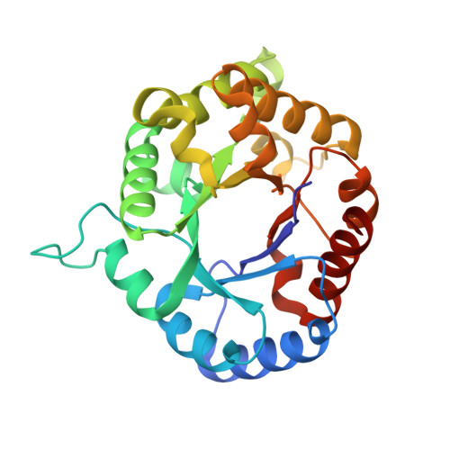
Triosephosphate isomerase is a common crystallization contaminant of soluble His-tagged proteins produced in Escherichia coli.

Kozlov, G.Vinaik, R.Gehring, K.

(2013) Acta Crystallogr Sect F Struct Biol Cryst Commun 69: 499-502

  • DOI: https://doi.org/10.1107/S1744309113010841
  • Primary Citation Related Structures: 
    4IOT

  • PubMed Abstract: 

    Attempts to crystallize several mammalian proteins overexpressed in Escherichia coli revealed a common contaminant, triosephosphate isomerase, a protein involved in glucose metabolism. Even with triosephosphate isomerase present in very small amounts, similarly shaped crystals appeared in the crystallization drops in a number of polyethylene glycol-containing conditions. All of the target proteins were His-tagged and their purification involved immobilized metal-affinity chromatography (IMAC), a step that was likely to lead to triosephosphate isomerase contamination. Analysis of the triosephosphate isomerase crystals led to the structure of E. coli triosephosphate isomerase at 1.85 Å resolution, which is a significant improvement over the previous structure.


  • Organizational Affiliation
    • Department of Biochemistry, Groupe de Recherche axé sur la Structure des Protéines, McGill University, 3649 Promenade Sir William Osler, Montréal, Québec H3G 0B1, Canada.

Macromolecule Content 

  • Total Structure Weight: 54.1 kDa 
  • Atom Count: 4,306 
  • Modeled Residue Count: 508 
  • Deposited Residue Count: 510 
  • Unique protein chains: 1

Macromolecules

Find similar proteins by:|  3D Structure
Entity ID: 1
MoleculeChains  Sequence LengthOrganismDetailsImage
Triosephosphate isomerase
A, B
255Escherichia coli str. K-12 substr. DH10BMutation(s): 0 
EC: 5.3.1.1
UniProt
Find proteins for B1XB85 (Escherichia coli (strain K12 / DH10B))
Explore B1XB85 
Go to UniProtKB:  B1XB85
Entity Groups
Sequence Clusters30% Identity50% Identity70% Identity90% Identity95% Identity100% Identity
UniProt GroupB1XB85
Sequence Annotations
Expand
Reference Sequence

Small Molecules

Ligands 1 Unique
IDChains Name / Formula / InChI Key2D Diagram3D Interactions
SO4

Query on SO4


Download:Ideal Coordinates CCD File
C [auth A]SULFATE ION
O4 S
QAOWNCQODCNURD-UHFFFAOYSA-L

Experimental Data & Validation

Experimental Data

  • Method: X-RAY DIFFRACTION
  • Resolution: 1.85 Å
  • R-Value Free:  0.206 (Depositor), 0.209 (DCC) 
  • R-Value Work:  0.172 (Depositor), 0.173 (DCC) 
  • R-Value Observed: 0.174 (Depositor) 
Space Group: P 21 21 21
Unit Cell:
Length ( Å )Angle ( ˚ )
a = 46.47α = 90
b = 67.452β = 90
c = 150.354γ = 90
Software Package:
Software NamePurpose
ADSCdata collection
PHASERphasing
REFMACrefinement
HKL-2000data reduction
HKL-2000data scaling

Structure Validation

View Full Validation Report


Entry History 

Deposition Data

Revision History  (Full details and data files)

  • Version 1.0: 2013-01-23
    Type: Initial release
  • Version 1.1: 2013-06-05
    Changes: Database references
  • Version 1.2: 2023-09-20
    Changes: Data collection, Database references, Derived calculations, Refinement description