4INC | pdb_00004inc

Human Histidine Triad Nucleotide Binding Protein 2


Experimental Data Snapshot

  • Method: X-RAY DIFFRACTION
  • Resolution: 1.19 Å
  • R-Value Free: 
    0.178 (Depositor), 0.179 (DCC) 
  • R-Value Work: 
    0.160 (Depositor), 0.161 (DCC) 
  • R-Value Observed: 
    0.161 (Depositor) 

Starting Model: experimental
View more details

wwPDB Validation 3D Report Full Report

Validation slider image for 4INC

This is version 1.2 of the entry. See complete history

Literature

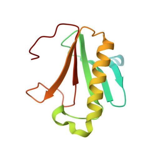
Structural characterization of human histidine triad nucleotide-binding protein 2, a member of the histidine triad superfamily.

Maize, K.M.Wagner, C.R.Finzel, B.C.

(2013) FEBS J 280: 3389-3398

  • DOI: https://doi.org/10.1111/febs.12330
  • Primary Citation Related Structures: 
    4INC, 4INI

  • PubMed Abstract: 

    The histidine triad proteins (HITs) constitute a large and ubiquitous superfamily of nucleotide hydrolases. The human histidine triad nucleotide-binding proteins (hHints) are a distinct class of HITs noted for their acyl-AMP hydrolase and phosphoramidase activity. The first high-resolution crystal structures of hHint2 with and without bound AMP are described. The differences between hHint2 and previously known HIT family protein structures are discussed. HIT family enzymes have historically been divided into five classes based on their catalytic specificity: Hint, fragile HIT protein, galactose-1-phosphate uridylyltransferase, DcpS and aprataxin. However, although several structures exist for the enzymes in these classes, the endogenous substrates of many of these enzymes have not been identified or biochemically characterized. To better understand the structural relationships of the HIT enzymes, a structure-based phylogeny was constructed that resulted in the identification of several new putative HIT clades with potential acyl-AMP hydrolase and phosphoramidase activity.


  • Organizational Affiliation
    • Department of Medicinal Chemistry, University of Minnesota, Minneapolis, MN, USA.

Macromolecule Content 

  • Total Structure Weight: 28.19 kDa 
  • Atom Count: 2,171 
  • Modeled Residue Count: 213 
  • Deposited Residue Count: 260 
  • Unique protein chains: 1

Macromolecules

Find similar proteins by:|  3D Structure
Entity ID: 1
MoleculeChains  Sequence LengthOrganismDetailsImage
Histidine triad nucleotide-binding protein 2, mitochondrial
A, B
130Homo sapiensMutation(s): 0 
Gene Names: HINT2
EC: 3 (PDB Primary Data), 3.9.1 (UniProt)
UniProt & NIH Common Fund Data Resources
Find proteins for Q9BX68 (Homo sapiens)
Explore Q9BX68 
Go to UniProtKB:  Q9BX68
PHAROS:  Q9BX68
GTEx:  ENSG00000137133 
Entity Groups
Sequence Clusters30% Identity50% Identity70% Identity90% Identity95% Identity100% Identity
UniProt GroupQ9BX68
Sequence Annotations
Expand
Reference Sequence

Small Molecules

Ligands 1 Unique
IDChains Name / Formula / InChI Key2D Diagram3D Interactions
CL

Query on CL


Download:Ideal Coordinates CCD File
C [auth A]CHLORIDE ION
Cl
VEXZGXHMUGYJMC-UHFFFAOYSA-M

Experimental Data & Validation

Experimental Data

  • Method: X-RAY DIFFRACTION
  • Resolution: 1.19 Å
  • R-Value Free:  0.178 (Depositor), 0.179 (DCC) 
  • R-Value Work:  0.160 (Depositor), 0.161 (DCC) 
  • R-Value Observed: 0.161 (Depositor) 
Space Group: P 21 21 21
Unit Cell:
Length ( Å )Angle ( ˚ )
a = 37.826α = 90
b = 74.952β = 90
c = 77.796γ = 90
Software Package:
Software NamePurpose
SCALAdata scaling
PHASERphasing
PHENIXrefinement
PDB_EXTRACTdata extraction
JDirectordata collection
XSCALEdata scaling

Structure Validation

View Full Validation Report


Entry History 

Deposition Data

Revision History  (Full details and data files)

  • Version 1.0: 2013-05-22
    Type: Initial release
  • Version 1.1: 2013-07-17
    Changes: Database references
  • Version 1.2: 2023-09-20
    Changes: Data collection, Database references, Derived calculations, Refinement description