4IN7 | pdb_00004in7

(M)L214N mutant of the Rhodobacter sphaeroides Reaction Center


Experimental Data Snapshot

  • Method: X-RAY DIFFRACTION
  • Resolution: 2.85 Å
  • R-Value Free: 
    0.213 (Depositor), 0.211 (DCC) 
  • R-Value Work: 
    0.174 (Depositor), 0.179 (DCC) 
  • R-Value Observed: 
    0.176 (Depositor) 

wwPDB Validation 3D Report Full Report

Validation slider image for 4IN7

Ligand Structure Quality Assessment 


This is version 1.2 of the entry. See complete history

Literature

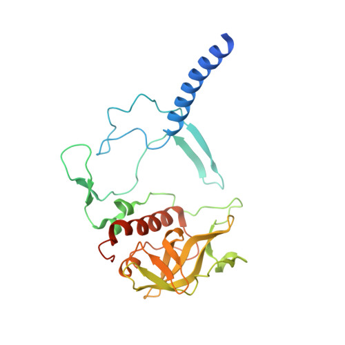
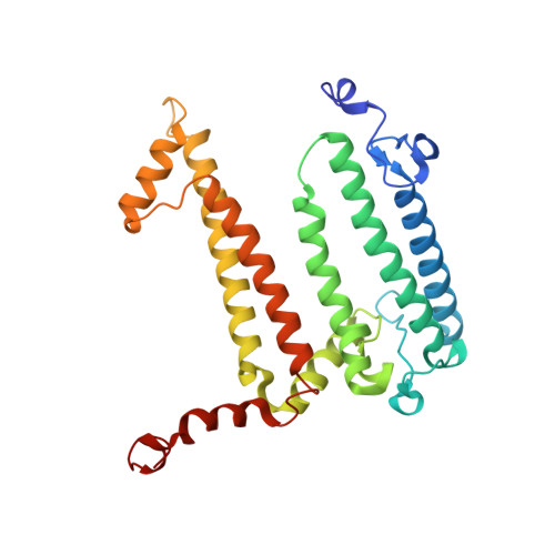
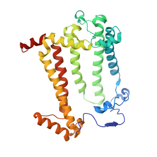
Role of Rhodobacter sphaeroides photosynthetic reaction center residue M214 in the composition, absorbance properties, and conformations of H(A) and B(A) cofactors.

Saer, R.G.Hardjasa, A.Rosell, F.I.Mauk, A.G.Murphy, M.E.Beatty, J.T.

(2013) Biochemistry 52: 2206-2217

  • DOI: https://doi.org/10.1021/bi400207m
  • Primary Citation Related Structures: 
    4IN5, 4IN6, 4IN7

  • PubMed Abstract: 

    In the native reaction center (RC) of Rhodobacter sphaeroides, the side chain of (M)L214 projects orthogonally toward the plane and into the center of the A branch bacteriopheophytin (BPhe) macrocycle. The possibility that this side chain is responsible for the dechelation of the central Mg(2+) of bacteriochlorophyll (BChl) was investigated by replacement of (M)214 with residues possessing small, nonpolar side chains that can neither coordinate nor block access to the central metal ion. The (M)L214 side chain was also replaced with Cys, Gln, and Asn to evaluate further the requirements for assembly of the RC with BChl in the HA pocket. Photoheterotrophic growth studies showed no difference in growth rates of the (M)214 nonpolar mutants at a low light intensity, but the growth of the amide-containing mutants was impaired. The absorbance spectra of purified RCs indicated that although absorbance changes are associated with the nonpolar mutations, the nonpolar mutant RC pigment compositions are the same as in the wild-type protein. Crystal structures of the (M)L214G, (M)L214A, and (M)L214N mutants were determined (determined to 2.2-2.85 Å resolution), confirming the presence of BPhe in the HA pocket and revealing alternative conformations of the phytyl tail of the accessory BChl in the BA site of these nonpolar mutants. Our results demonstrate that (i) BChl is converted to BPhe in a manner independent of the aliphatic side chain length of nonpolar residues replacing (M)214, (ii) BChl replaces BPhe if residue (M)214 has an amide-bearing side chain, (iii) (M)214 side chains containing sulfur are not sufficient to bind BChl in the HA pocket, and (iv) the (M)214 side chain influences the conformation of the phytyl tail of the BA BChl.


  • Organizational Affiliation
    • Department of Microbiology and Immunology, The University of British Columbia, 2350 Health Sciences Mall, Vancouver, BC, Canada V6T 1Z3.

Macromolecule Content 

  • Total Structure Weight: 108.03 kDa 
  • Atom Count: 7,359 
  • Modeled Residue Count: 823 
  • Deposited Residue Count: 855 
  • Unique protein chains: 3

Macromolecules

Find similar proteins by:|  3D Structure
Entity ID: 1
MoleculeChains  Sequence LengthOrganismDetailsImage
Reaction center protein H chainA [auth H]266Cereibacter sphaeroidesMutation(s): 0 
Gene Names: puhA
Membrane Entity: Yes 
UniProt
Find proteins for P0C0Y7 (Cereibacter sphaeroides)
Explore P0C0Y7 
Go to UniProtKB:  P0C0Y7
Entity Groups
Sequence Clusters30% Identity50% Identity70% Identity90% Identity95% Identity100% Identity
UniProt GroupP0C0Y7
Sequence Annotations
Expand
Reference Sequence
Find similar proteins by:|  3D Structure
Entity ID: 2
MoleculeChains  Sequence LengthOrganismDetailsImage
Reaction center protein L chainB [auth L]282Cereibacter sphaeroidesMutation(s): 0 
Gene Names: pufL
Membrane Entity: Yes 
UniProt
Find proteins for P0C0Y8 (Cereibacter sphaeroides)
Explore P0C0Y8 
Go to UniProtKB:  P0C0Y8
Entity Groups
Sequence Clusters30% Identity50% Identity70% Identity90% Identity95% Identity100% Identity
UniProt GroupP0C0Y8
Sequence Annotations
Expand
Reference Sequence
Find similar proteins by:|  3D Structure
Entity ID: 3
MoleculeChains  Sequence LengthOrganismDetailsImage
Reaction center protein M chainC [auth M]307Cereibacter sphaeroidesMutation(s): 1 
Gene Names: pufM
Membrane Entity: Yes 
UniProt
Find proteins for P0C0Y9 (Cereibacter sphaeroides)
Explore P0C0Y9 
Go to UniProtKB:  P0C0Y9
Entity Groups
Sequence Clusters30% Identity50% Identity70% Identity90% Identity95% Identity100% Identity
UniProt GroupP0C0Y9
Sequence Annotations
Expand
Reference Sequence

Small Molecules

Ligands 14 Unique
IDChains Name / Formula / InChI Key2D Diagram3D Interactions
CDL

Query on CDL


Download:Ideal Coordinates CCD File
DA [auth M]CARDIOLIPIN
C81 H156 O17 P2
XVTUQDWPJJBEHJ-KZCWQMDCSA-L
GGD

Query on GGD


Download:Ideal Coordinates CCD File
J [auth H]NONADEC-10-ENOIC ACID 2-[3,4-DIHYDROXY-6-HYDROXYMETHYL-5-(3,4,5-TRIHYDROXY-6-HYDROXYMETHYL-TETRAHYDRO-PYRAN-2-YLOXY)-TETRAHYDRO-PYRAN-2-YLOXY] -1-OCTADEC-9-ENOYLOXYMETHYL-ETHYL ESTER
C52 H94 O15
OGKQOSUHVSQNSH-IQXSWRQFSA-N
BCL

Query on BCL


Download:Ideal Coordinates CCD File
K [auth L],
P [auth L],
V [auth M],
W [auth M]
BACTERIOCHLOROPHYLL A
C55 H74 Mg N4 O6
DSJXIQQMORJERS-AGGZHOMASA-M
BPH

Query on BPH


Download:Ideal Coordinates CCD File
AA [auth M],
GA [auth M]
BACTERIOPHEOPHYTIN A
C55 H76 N4 O6
KWOZSBGNAHVCKG-SZQBJALDSA-N
U10

Query on U10


Download:Ideal Coordinates CCD File
BA [auth M],
O [auth L]
UBIQUINONE-10
C59 H90 O4
ACTIUHUUMQJHFO-UPTCCGCDSA-N
PC1

Query on PC1


Download:Ideal Coordinates CCD File
EA [auth M]1,2-DIACYL-SN-GLYCERO-3-PHOSPHOCHOLINE
C44 H88 N O8 P
NRJAVPSFFCBXDT-HUESYALOSA-N
SPO

Query on SPO


Download:Ideal Coordinates CCD File
CA [auth M]SPHEROIDENE
C41 H60 O
FJOCMTHZSURUFA-KXCOHNEYSA-N
LDA

Query on LDA


Download:Ideal Coordinates CCD File
L,
M [auth L],
N [auth L],
X [auth M],
Y [auth M]
LAURYL DIMETHYLAMINE-N-OXIDE
C14 H31 N O
SYELZBGXAIXKHU-UHFFFAOYSA-N
HTO

Query on HTO


Download:Ideal Coordinates CCD File
R [auth L],
S [auth L]
HEPTANE-1,2,3-TRIOL
C7 H16 O3
HXYCHJFUBNTKQR-RNFRBKRXSA-N
PO4

Query on PO4


Download:Ideal Coordinates CCD File
Q [auth L]PHOSPHATE ION
O4 P
NBIIXXVUZAFLBC-UHFFFAOYSA-K
GOL

Query on GOL


Download:Ideal Coordinates CCD File
D [auth H]
E [auth H]
F [auth H]
G [auth H]
H
D [auth H],
E [auth H],
F [auth H],
G [auth H],
H,
T [auth L],
U [auth L]
GLYCEROL
C3 H8 O3
PEDCQBHIVMGVHV-UHFFFAOYSA-N
FE

Query on FE


Download:Ideal Coordinates CCD File
Z [auth M]FE (III) ION
Fe
VTLYFUHAOXGGBS-UHFFFAOYSA-N
K

Query on K


Download:Ideal Coordinates CCD File
I [auth H]POTASSIUM ION
K
NPYPAHLBTDXSSS-UHFFFAOYSA-N
MG

Query on MG


Download:Ideal Coordinates CCD File
FA [auth M]MAGNESIUM ION
Mg
JLVVSXFLKOJNIY-UHFFFAOYSA-N

Experimental Data & Validation

Experimental Data

  • Method: X-RAY DIFFRACTION
  • Resolution: 2.85 Å
  • R-Value Free:  0.213 (Depositor), 0.211 (DCC) 
  • R-Value Work:  0.174 (Depositor), 0.179 (DCC) 
  • R-Value Observed: 0.176 (Depositor) 
Space Group: P 31 2 1
Unit Cell:
Length ( Å )Angle ( ˚ )
a = 139.138α = 90
b = 139.138β = 90
c = 185.14γ = 120
Software Package:
Software NamePurpose
SCALAdata scaling
REFMACrefinement
PDB_EXTRACTdata extraction
XDSdata scaling
XSCALEdata scaling

Structure Validation

View Full Validation Report


Ligand Structure Quality Assessment 


Entry History 

Deposition Data

Revision History  (Full details and data files)

  • Version 1.0: 2013-06-05
    Type: Initial release
  • Version 1.1: 2017-11-15
    Changes: Refinement description
  • Version 1.2: 2024-02-28
    Changes: Data collection, Database references, Derived calculations, Structure summary