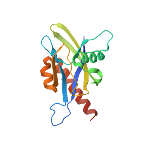
4IJX | pdb_00004ijx

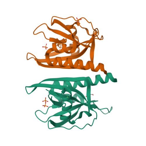
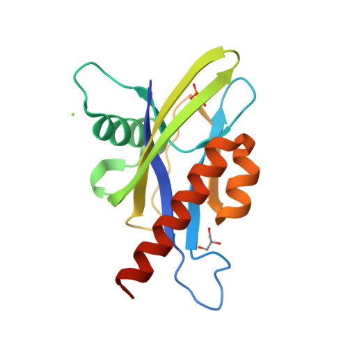
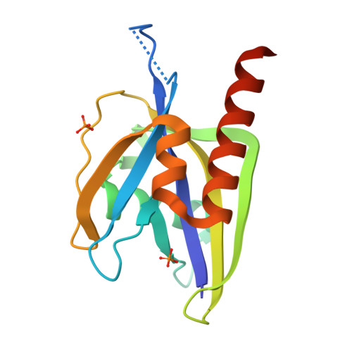
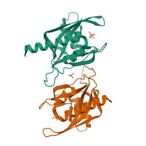
Crystal structure of human Ap4A hydrolase E58A mutant complexed with DPO


Experimental Data Snapshot

  • Method: X-RAY DIFFRACTION
  • Resolution: 2.10 Å
  • R-Value Free: 
    0.232 (Depositor), 0.230 (DCC) 
  • R-Value Work: 
    0.189 (Depositor), 0.190 (DCC) 
  • R-Value Observed: 
    0.191 (Depositor) 

Starting Model: experimental
View more details

wwPDB Validation   3D Report Full Report


Ligand Structure Quality Assessment 

Created with Raphaël 2.3.0Worse 01 BetterLigand structure goodness of fit to experimental dataBest fitted DPOClick on this verticalbar to view details

This is version 1.1 of the entry. See complete history


Literature

Crystal structure of wild-type and mutant human Ap4A hydrolase.

Ge, H.Chen, X.Yang, W.Niu, L.Teng, M.

(2013) Biochem Biophys Res Commun 432: 16-21

  • DOI: https://doi.org/10.1016/j.bbrc.2013.01.095
  • Primary Citation of Related Structures:  
    3U53, 4ICK, 4IJX

  • PubMed Abstract: 

    Ap4A hydrolase (asymmetrical diadenosine tetraphosphate hydrolase, EC 3.6.1.17), an enzyme involved in a number of biological processes, is characterized as cleaving the polyphosphate chain at the fourth phosphate from the bound adenosine moiety. This paper presents the crystal structure of wild-type and E58A mutant human Ap4A hydrolase. Similar to the canonical Nudix fold, human Ap4A hydrolase shows the common αβα-sandwich architecture. Interestingly, two sulfate ions and one diphosphate coordinated with some conserved residues were observed in the active cleft, which affords a better understanding of a possible mode of substrate binding.


  • Organizational Affiliation

    Institute of Health Sciences and Modern Experiment Technology Center, Anhui University, Hefei 230601, People's Republic of China. hhge@ahu.edu.cn


Macromolecules
Find similar proteins by:  (by identity cutoff)  |  3D Structure
Entity ID: 1
MoleculeChains Sequence LengthOrganismDetailsImage
Bis(5'-nucleosyl)-tetraphosphatase [asymmetrical]
A, B
155Homo sapiensMutation(s): 1 
Gene Names: NUDT2APAH1
EC: 3.6.1.17
UniProt & NIH Common Fund Data Resources
Find proteins for P50583 (Homo sapiens)
Explore P50583 
Go to UniProtKB:  P50583
PHAROS:  P50583
GTEx:  ENSG00000164978 
Entity Groups  
Sequence Clusters30% Identity50% Identity70% Identity90% Identity95% Identity100% Identity
UniProt GroupP50583
Sequence Annotations
Expand
  • Reference Sequence
Experimental Data & Validation

Experimental Data

  • Method: X-RAY DIFFRACTION
  • Resolution: 2.10 Å
  • R-Value Free:  0.232 (Depositor), 0.230 (DCC) 
  • R-Value Work:  0.189 (Depositor), 0.190 (DCC) 
  • R-Value Observed: 0.191 (Depositor) 
Space Group: P 43 21 2
Unit Cell:
Length ( Å )Angle ( ˚ )
a = 72.369α = 90
b = 72.369β = 90
c = 133.379γ = 90
Software Package:
Software NamePurpose
REFMACrefinement
HKL-2000data scaling

Structure Validation

View Full Validation Report


Ligand Structure Quality Assessment 

Created with Raphaël 2.3.0Worse 01 BetterLigand structure goodness of fit to experimental dataBest fitted DPOClick on this verticalbar to view details

Entry History 

Deposition Data

Revision History  (Full details and data files)

  • Version 1.0: 2013-03-20
    Type: Initial release
  • Version 1.1: 2023-11-08
    Changes: Data collection, Database references, Derived calculations, Refinement description