4II0 | pdb_00004ii0

Crystal structure of CrataBL, a trypsin inhibitor from Crataeva tapia


Experimental Data Snapshot

  • Method: X-RAY DIFFRACTION
  • Resolution: 1.75 Å
  • R-Value Free: 
    0.232 (Depositor), 0.230 (DCC) 
  • R-Value Work: 
    0.185 (Depositor), 0.185 (DCC) 
  • R-Value Observed: 
    0.186 (Depositor) 

wwPDB Validation 3D Report Full Report

Validation slider image for 4II0

Ligand Structure Quality Assessment 


This is version 2.1 of the entry. See complete history

Literature

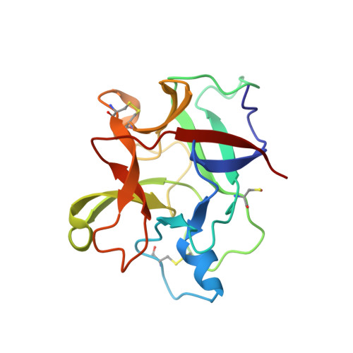
Crystal Structure of Crataeva tapia Bark Protein (CrataBL) and Its Effect in Human Prostate Cancer Cell Lines.

Ferreira, R.D.Zhou, D.Ferreira, J.G.Silva, M.C.Silva-Lucca, R.A.Mentele, R.Paredes-Gamero, E.J.Bertolin, T.C.Dos Santos Correia, M.T.Paiva, P.M.Gustchina, A.Wlodawer, A.Oliva, M.L.

(2013) PLoS One 8: e64426-e64426

  • DOI: https://doi.org/10.1371/journal.pone.0064426
  • Primary Citation Related Structures: 
    4IHZ, 4II0

  • PubMed Abstract: 

    A protein isolated from the bark of Crataeva tapia (CrataBL) is both a Kunitz-type plant protease inhibitor and a lectin. We have determined the amino acid sequence and three-dimensional structure of CrataBL, as well as characterized its selected biochemical and biological properties. We found two different isoforms of CrataBL isolated from the original source, differing in positions 31 (Pro/Leu); 92 (Ser/Leu); 93 (Ile/Thr); 95 (Arg/Gly) and 97 (Leu/Ser). CrataBL showed relatively weak inhibitory activity against trypsin (Kiapp = 43 µM) and was more potent against Factor Xa (Kiapp = 8.6 µM), but was not active against a number of other proteases. We have confirmed that CrataBL contains two glycosylation sites and forms a dimer at high concentration. The high-resolution crystal structures of two different crystal forms of isoform II verified the β-trefoil fold of CrataBL and have shown the presence of dimers consisting of two almost identical molecules making extensive contacts (∼645 Å(2)). The structure differs from those of the most closely related proteins by the lack of the N-terminal β-hairpin. In experiments aimed at investigating the biological properties of CrataBL, we have shown that addition of 40 µM of the protein for 48 h caused maximum growth inhibition in MTT assay (47% of DU145 cells and 43% of PC3 cells). The apoptosis of DU145 and PC3 cell lines was confirmed by flow cytometry using Annexin V/FITC and propidium iodide staining. Treatment with CrataBL resulted in the release of mitochondrial cytochrome c and in the activation of caspase-3 in DU145 and PC3 cells.


  • Organizational Affiliation
    • Departamento de Bioquímica, Universidade Federal de São Paulo, São Paulo, São Paulo, Brazil.

Macromolecule Content 

  • Total Structure Weight: 39.05 kDa 
  • Atom Count: 3,027 
  • Modeled Residue Count: 328 
  • Deposited Residue Count: 330 
  • Unique protein chains: 1

Macromolecules

Find similar proteins by:|  3D Structure
Entity ID: 1
MoleculeChains  Sequence LengthOrganismDetailsImage
CrataBL
A, B
165Crateva tapiaMutation(s): 0 
UniProt
Find proteins for U3KRG0 (Crateva tapia)
Explore U3KRG0 
Go to UniProtKB:  U3KRG0
Entity Groups
Sequence Clusters30% Identity50% Identity70% Identity90% Identity95% Identity100% Identity
UniProt GroupU3KRG0
Glycosylation
Glycosylation Sites: 2
Sequence Annotations
Expand
Reference Sequence

Oligosaccharides

Help  
Entity ID: 2
MoleculeChains Length2D Diagram GlycosylationD Interactions
alpha-L-fucopyranose-(1-3)-[2-acetamido-2-deoxy-beta-D-glucopyranose-(1-4)]2-acetamido-2-deoxy-beta-D-glucopyranose
C
3N-Glycosylation
Glycosylation Resources
GlyTouCan: G11971MR
GlyCosmos: G11971MR
GlyGen: G11971MR

Small Molecules

Ligands 3 Unique
IDChains Name / Formula / InChI Key2D Diagram3D Interactions
NAG

Query on NAG


Download:Ideal Coordinates CCD File
K [auth A],
S [auth B],
T [auth B]
2-acetamido-2-deoxy-beta-D-glucopyranose
C8 H15 N O6
OVRNDRQMDRJTHS-FMDGEEDCSA-N
SO4

Query on SO4


Download:Ideal Coordinates CCD File
D [auth A]
E [auth A]
F [auth A]
G [auth A]
H [auth A]
D [auth A],
E [auth A],
F [auth A],
G [auth A],
H [auth A],
L [auth B],
M [auth B],
N [auth B],
O [auth B],
P [auth B]
SULFATE ION
O4 S
QAOWNCQODCNURD-UHFFFAOYSA-L
GOL

Query on GOL


Download:Ideal Coordinates CCD File
I [auth A],
J [auth A],
Q [auth B],
R [auth B]
GLYCEROL
C3 H8 O3
PEDCQBHIVMGVHV-UHFFFAOYSA-N
Modified Residues  1 Unique
IDChains TypeFormula2D DiagramParent
CSX
Query on CSX
A, B
L-PEPTIDE LINKINGC3 H7 N O3 SCYS

Experimental Data & Validation

Experimental Data

  • Method: X-RAY DIFFRACTION
  • Resolution: 1.75 Å
  • R-Value Free:  0.232 (Depositor), 0.230 (DCC) 
  • R-Value Work:  0.185 (Depositor), 0.185 (DCC) 
  • R-Value Observed: 0.186 (Depositor) 
Space Group: C 1 2 1
Unit Cell:
Length ( Å )Angle ( ˚ )
a = 95.585α = 90
b = 76.279β = 120.08
c = 62.335γ = 90
Software Package:
Software NamePurpose
HKL-2000data collection
PHASESphasing
REFMACrefinement
HKL-3000data reduction
SCALEPACKdata scaling

Structure Validation

View Full Validation Report


Ligand Structure Quality Assessment 


Entry History 

Deposition Data

Revision History  (Full details and data files)

  • Version 1.0: 2013-07-24
    Type: Initial release
  • Version 2.0: 2020-07-29
    Type: Remediation
    Reason: Carbohydrate remediation
    Changes: Atomic model, Data collection, Derived calculations, Structure summary
  • Version 2.1: 2025-03-26
    Changes: Data collection, Database references, Structure summary